Image source: NNUH
News • Reducing collateral damage
A shield to protect patients during prostate cancer radiotherapy
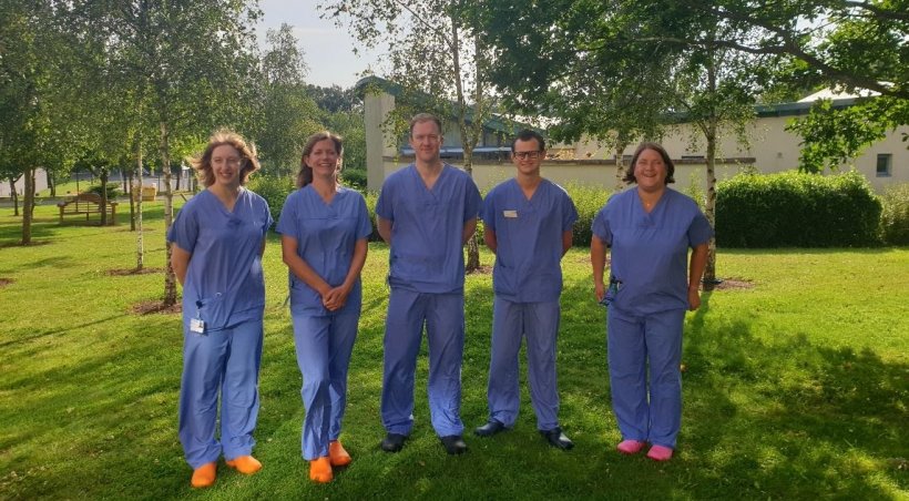
Prostate cancer specialists from the Radiotherapy Department at Norfolk and Norwich University Hospitals Foundation Trust have become the first in the world to use an innovative technique to help patients receiving treatment for prostate cancer.
Some patients receiving radiotherapy for prostate cancer will have their treatment split into two portions. The first stage of killing the cancerous cells uses a temporary radioactive implant, in a process known as high dose rate (HDR) brachytherapy. The second part is delivered as a powerful x-ray beam from outside the patient, in a process known as external beam radiotherapy, which is carried out over a number of appointments. During both stages, however, it is possible for healthy tissue to be damaged such as the large bowel which can become chronically inflamed.
By inserting a special “shield” known as a hyaluronic acid rectal spacer, it is possible to protect the neighbouring tissues from the potential damage caused by external beam radiotherapy. The rectal spacer insertion is usually carried out under local anaesthetic, one to two weeks prior to treatment. However, in June the Brachytherapy Team became the first team globally to insert a hyaluronic acid rectal spacer during HDR brachytherapy treatment.
The insertion of the implant immediately before an HDR procedure can not only reduce the number of appointments a patient has to have, but can also enable the treatment team to plan in real time and better position needles
Jenny Nobes
The composition of other spacing devices has prevented their use during HDR brachytherapy treatment, as they limit the visibility of the ultrasound imaging, which is key for monitoring this type of brachytherapy treatment. The hyaluronic acid spacer does not interfere with the ultrasound signals which means the prostate gland and surrounding organs can be seen fully after the implant has been inserted. This allows the implant to be inserted during the HDR procedure without reducing image quality for the radiotherapist placing the needle.
NNUH Consultant Clinical Oncologist Dr Jenny Nobes, said: “This is great news for our patients as it reduces the amount of tissue damage to the surrounding area, potentially reducing side effects and reduces the amount of time they have to spend in the hospital. The insertion of the implant immediately before an HDR procedure can not only reduce the number of appointments a patient has to have, but can also enable the treatment team to plan in real time and better position needles, which again is better for our patients.”
Source: Norfolk and Norwich University Hospitals Foundation Trust
25.08.2021


