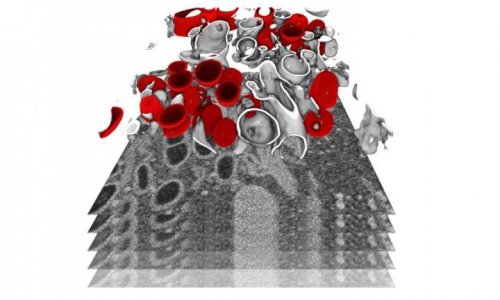
Image source: Umeå University/Selma Dahmane
News • Cryo-electron tomography
3D microscopy reveals how tick-borne virus replicates
Researchers at Umeå University show how tick‑borne viruses remodel human cells into virus factories, using an advanced microscopy method.
The findings provide new insight into how the virus replicates and matures, knowledge that may become important for future treatments against TBE. The study is published in Nature Communications.

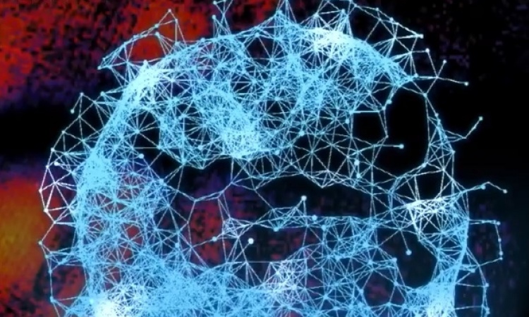
Image source: Umeå University/Mattias Pettersson
“When we saw the three‑dimensional images for the first time, we immediately realized how much new information we could gain about the virus’s replication,” says Lars‑Anders Carlson, professor at the Department of Medical Chemistry and Biophysics at Umeå University, who led the study.
One of the most dangerous viral diseases spread in Europe is tick‑borne encephalitis. A bite from an infected tick can transmit the TBE virus to humans and cause severe inflammation of the brain. Using electron microscopy, researchers at Umeå University have now discovered how tick‑borne viruses reshape infected human cells and turn them into virus factories.
“It has been difficult to conduct this type of study on the TBE virus because it is so dangerous that we are not allowed to work with it at the electron microscope. But we managed to use a closely related virus, Langat virus, which behaves almost identically in cells but is far less dangerous to humans. Both belong to the flavivirus genus,” explains Lars‑Anders Carlson.
With cryo‑electron tomography, a specialized form of electron microscopy, the researchers were able to create detailed three‑dimensional images of the interior of infected cells that were rapidly frozen and preserved in a life‑like state. This revealed how the virus r the interior of the cell to create the perfect environment for hiding the mass production of viral genes.

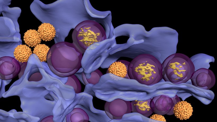
Image source: Umeå University/Selma Dahmane
The researchers could also show how new virus particles are produced right next to the viral “gene factories,” and how these new particles change shape from an “immature” form to the mature form that is then released from the cells. By comparing two different variants of the virus, they further observed how a very small genetic difference between them led to different maturation speeds.
“Here we were able to directly observe how a small change in a single gene caused the virus to mature at different rates,” says Bina Singh, postdoctoral researcher at the Department of Medical Chemistry and Biophysics at Umeå University. Achieving this level of detailed understanding requires more than advanced technology. Research of this kind depends on long‑term resources, the right expertise, and close collaboration among many skilled scientists. These factors were crucial for the project’s development from an initiative based in Umeå to a broad international collaboration.
The project began with funding from the Umeå Centre for Microbial Research (UCMR), which brings together infection biology researchers at Umeå University. “Their postdoctoral programme ‘Excellence by Choice’ made it possible to recruit two talented international researchers to Umeå: Jianguo Zhang and Erin Schexnaydre,” says Lars-Anders Carlson.
In the groups of Lars‑Anders Carlson and Anna Överby, and in close collaboration with the Umeå Centre for Electron Microscopy (UCEM), Jianguo Zhang and Erin Schexnaydre developed ambitious new methods for cryo‑electron tomography of tick‑borne viruses in infected cells and mouse brains.
The completion of the study was made possible through expanded collaboration with research colleagues in Norway and the United States, funded by major, collaboration‑focused grants from the Swedish Research Council and the Knut and Alice Wallenberg Foundation.
Source: Umeå University; text: Ingrid Söderbergh
09.04.2026