Validation of the value of cardiovascular MR
Research results will be presented and discussed at ECR 2009
After two years of intensive work the results from the German pilot phase of the EuroCMR Register are due to be published in the forthcoming issue of the Journal of the American College of Cardiology*, and also presented and discussed in detail at this year's ECR in Barcelona.
From April 2007 to January 2009, cardiologists from six countries, led by Dr Oliver Bruder of the Department of Cardiology and Angiology, Elisabeth Hospital, Essen, and Dr Heiko Mahrholdt, at the Department of Cardiology, Robert Bosch Medical Centre, Stuttgart, have been evaluating data from 11,040 patients. The aim has been to prove the relevance of magnetic resonance imaging (MRI) in cardiac diagnostics. The results will present the first valid data on indication, image quality, safety and benefit for patient management of CMR (Cardiovascular Magnetic Resonance) to the world of cardiology.
They report that CMR has proved to be a safe procedure for the diagnosis of myocarditis/cardiomyocarditis; risk stratification for suspected diseases of the coronary arties (ischaemia) and to assess a patient’s chances of survival. In 98% of examinations the diagnostic image quality achieved impacted on patient management significantly.
The findings have attracted considerable interest, not least because they show that precise cardiac diagnostics can be achieved non-invasively and without damaging radiation exposure.
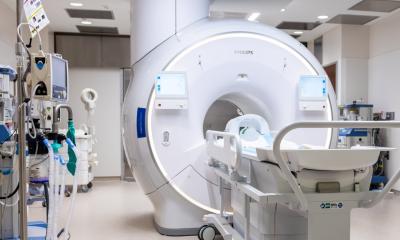
Cardiovascular magnetic resonance (CMR) is a rapidly emerging non-invasive imaging technique providing high-resolution images of the heart in any desired plane without application of radiation. Rather than a single technique, CMR consists of several protocols that can be performed in various combinations during a single examination. For example, cine-CMR can provide cardiac morphology, function and contractile reserve; perfusion CMR, with and without vasodilators, can provide myocardial perfusion, and contrast CMR can be used for infarct detection, as well as for tissue characterisation.
CMR has a broad range of appropriate clinical applications and is increasingly used in daily clinical practice. However, detailed information on the general use of this technique in clinical routine, its safety and impact on patient management is currently not available. Thus, the German pilot phase of the EuroCMR (European Cardiovascular Magnetic Resonance) registry sought to evaluate indications, image quality, safety, and impact on patient management of routine CMR imaging in a large number of cases to:
• substantiate the clinical value of CMR
• help define clinical questions to be investigated as specific protocols on a European multi-centre level
For additional information from a US point of view, please click here .
* EuroCMR (European Cardiovascular Magnetic Resonance) Registry: Results of the German Pilot Phase. Oliver Bruder, Steffen Schneider, Detlef Nothnagel, Thorsten Dill, Vinzenz Hombach, Jeanette Schulz-Menger, Eike Nagel, Massimo Lombardi, Albert C. von Rossum, Anja Wagner, Juerg Schwitter, Jochen Senges, Georg V. Sabin, Udo Sechtem, and Heiko Marholdt. J. Am. Coll. Cardiol. Published online, 12 August 2009; doi: 10.1016/j.jacc.2009.07.003
01.09.2009


