Interventional radiology
It's diversified but precise
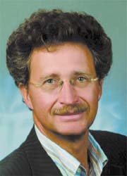
Along with paediatric radiology, interventional radiology will have a high profile at the 89th German Radiology Congress and 5th Joint Congress with the Austrian Radiology Society. During a discussion with Meike Lerner of European Hospital congress president Professor Dierk Vorwerk outlined what's on the agenda for the expected 6,900 visitors.
Training, he pointed out, will aim at those preparing to specialise in radiology. ‘In addition, we’d like to familiarise non-radiologists (e.g. general practitioners) with radiological results.’
International exchange will also be promoted; guests at this year’s congress will come from France, Korea and Turkey.
Asked about recent developments in interventional radiology, Prof. Vorwerk observed that the field is split into two areas: vascular and non-vascular intervention. ‘In the first, there has been particular progress in therapy for vessels of the lower leg. We can now intervene right down to the ankle. Initially, the problem was how to cover the long distance with a catheter, and also the vessels are very thin there. New developments now allow us to treat these indications with a high rate of success and, as the case may be, prevent amputation.
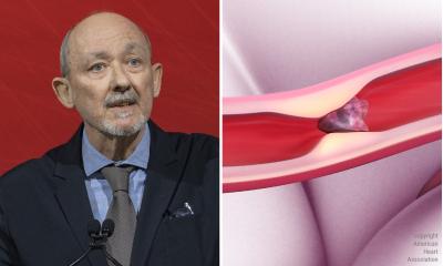
‘Developments in neuro-intervention in the invasive therapy for strokes, where we can significantly reduce the severity of an event, are also worth particular mention. There have always been efforts to keep the damaged brain areas as small as possible, which used to be done exclusively with medication – requiring a certain amount of treatment time. Now, we have mechanical procedures, intravascular and angiographic, to reduce a thrombus. Interventional therapy is usually combined with subsequent lysis therapy. Therefore, we can treat a stroke much faster and, as the case may be, more successfully. All in all these innovations in the vascular field are based on the development of new, more stable and thinner materials,’ he concluded.
Interventional oncology
In the non-vascular field, another topic of interest is interventional oncology, which is also partly carried out through the vessels. ‘In interventional oncology there is no surgery, or systemic chemotherapy to shrink and destroy a tumour. Currently, this therapy is focused on the liver, but increasingly also on the lungs, kidney and adrenal gland. Tumours on these organs are treated with physical media, with access through the skin. What we mean by physical media are particularly heat and cold; both can destroy tumours. We generate heat through microwaves, but primarily through radiofrequency ablation.
‘This treatment can be combined with medical therapy, where small “submarines” take chemotherapeutics to tumour locations, so systemic chemotherapy is not needed, and the effect is exclusively directed at a tumour. We can also treat a tumour radioactively in this way, so that its routes of supply are interrupted and the carcinoma is destroyed through radiation. This is called SIRT therapy, which is now covered by medical insurers.
‘With these procedures it remains to be seen how often they can be used and which kinds of tumour should be treated. Several studies are aiming to find answers to these questions.’
At the congress, the professor said radiologists and vascular surgeons will discuss ways to optimise vascular disease therapies, e.g. by setting up vascular treatment centres. ‘As radiologists we see our main task is in the diagnosis of these diseases and in providing minimally invasive therapy. Surgeons undertake difficult vascular surgery, which is becoming increasingly challenging. Vascular surgery must keep up with the increasing demand, and meet it comprehensively. The entire field of interventional radiology will remain exciting and the congress offers a forum where the current state of affairs and future developments will be discussed.’
30.04.2008


