Virtual Touch Tissue Imaging and Quantification
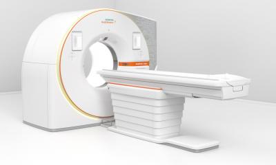
Acoustic Radiation Force Impulse Imaging (ARFI) is a new tissue strain imaging technology that utilizes sound waves to interrogate the mechanical stiffness properties of tissue. Virtual Touch™ Tissue Imaging and Virtual Touch™ Tissue Quantification are available on the Siemens ACUSON S2000™ ultrasound system.
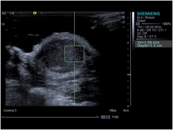
(Vs=1,58 m/s). This value is related to tissue stiffness at the user-defined anatomical region
localised using a conventional ultrasound image
These two innovative applications provide numerical and visual information on tissue stiffness:
Virtual Touch Tissue Quantification is the first application to provide a numerical value of shear-wave speed related to tissue stiffness at a precise anatomical location. Acoustic short duration pulses are emitted into the tissue. The impulses induce localized displacements within the tissue which allow shear waves to spread perpendicular to the excitation beam. The shear wave velocity (Vs) can be detected with ultrasonic waves, is measured in m/s and will be higher in stiffer tissues (Fig. 1).
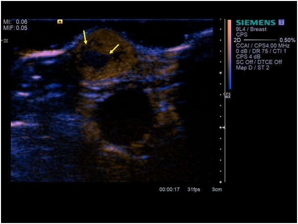
The Virtual Touch Image is a qualitative grayscale map of relative tissue stiffness in a user defined region of interest (ROI). This information is computed by examining the relative displacements of tissue elements due to an acoustic push pulse sent by the probe. In the Virtual Touch Image, bright regions depict tissue that is more elastic (less stiff) than dark regions (more stiff) (Fig. 2 and 3).
In several studies the diagnostic value of this new technique has been applied successfully in liver and kidney diseases [1-2].
In the past the limitation of this new technique was that this technique only works on low frequency probes. Due to the results of several ARFI studies and the demand of the user, Siemens has implemented this technique on a higher frequency probe. For the first time the user will now be able to use the high density Virtual Touch Tissue Imaging and Quantification as new diagnostic tool in diseases of the superficial organs like breast, thyroid, lymph nodes and testis.
These first results seem to be very interesting in different diseases and the technique even works in small lesions with a diameter less than 10 mm.
This marks an important step in a development that will redefine how we use ultrasound in the diagnosis, treatment, and therapy of superficial diseases.
02.03.2010