The future of digital mammography
By R Schulz-Wendtland, Erlangen University Hospital, Radiology Institute; T Wacker, Erlangen-Nurnberg University, and K-P Hermann, Department for Diagnostic Radiology, Gottingen University Hospital
Digital mammography, which is considered the gold standard of breast imaging, has rendered image management and consequently the entire – purely on-screen – diagnostic process more reliable and more efficient.
It is, however, the most recent developments affecting the entire digital system that promise to realise the full innovative potential of digital mammography for the benefit of patients. The most advanced of these technologies – which is already in use – is the so-called computer-aided detection (CAD) to review digital mammograms. Today, CAD applied as a second reader can help to improve the experienced reviewer’s specificity. State-of-the-art CAD systems built on the concept of neuronal networks can continuously increase specificity since they automatically integrate histological verifications of detected results in a database for future comparison with new mammograms. Such learning CAD systems are currently undergoing scientific tests.
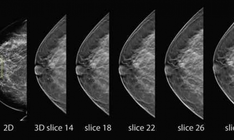
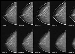
Currently, digital breast tomosynthesis (DBT) (Fig. 1) seems to be the most important innovation in mammography. DBT opens the door towards 3-D mammography and provides new possibilities for improving size and volume determination of findings which will be increasingly relevant for therapy purposes (Fig. 2a,b,c).
The different approaches towards contrast-enhanced digital mammography are interesting, particularly with regard to dense breasts and multi-focal and multi-centre findings. However, the lack of indications prevents standardised use of this technique. Moreover: Since this modality requires additional radiation exposure it will have a hard time competing with contrast-enhanced MR mammography, which is internationally accepted and does not require radiation.
In principle, computed tomography (CT) could also provide 3-D images of the breast. A CT system specifically developed for breast diagnostics is currently being tested clinically and compared to a conventional mammography system. With regard to the detection of nodules (masses) the CT system was superior to the conventional film-based mammography system. However, the latter yielded better results with regard to the visualisation of micro-calcifications.
Hybrid systems – the way to go?
A digital mammography system is the ideal platform to integrate other modalities such as ultrasound including elastography as well as alternative methods such as optical imaging or electrical impedance. With integrated modalities, different examinations can be performed almost parallel and provide 3-D data sets – the foundation of a promising image fusion.
For some years now, several manufacturers have been working on the integration of digital mammography and ultrasound with the aim to combine X-ray mammography’s high sensitivity with sonography’s high specificity. At the same time attempts are being made to automate ultrasound examinations in order to render them as operator-independent as possible (hybrid systems).
A digital tomosynthesis system will most likely form the basis for any future digital mammography equipment.
In the foreseeable future planar mammograms will be replaced by volume imaging / tumour targeting which benefit from the possibility to fuse images from different modalities (Fig. 3a,b,c). At the same time, computer-aided detection will improve due to these 3-D data sets. It was digital mammography that paved the way for these advancements.
22.07.2009