Video • Cell coordination explored
Why wound-healing comes in waves
How do cells in our bodies ask for directions? Without any maps to guide them, they still know where to go to heal wounds and renew our bodies.
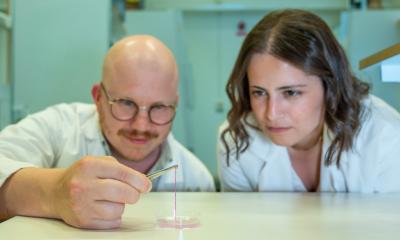
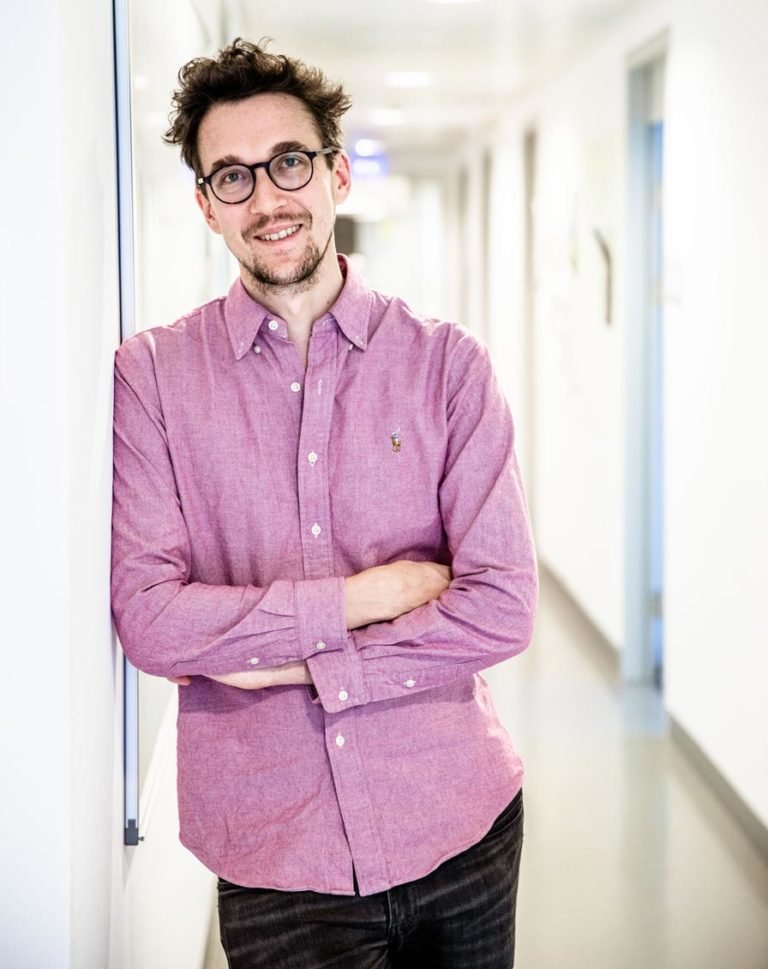
Edouard Hannezo and his group at the Institute of Science and Technology Austria (IST Austria) together with Tsuyoshi Hirashima and his group at Kyoto University just published a new study in Nature Physics that shows how mechanical and chemical waves coordinate the movement of cells.
© IST Austria
The question of how cells location of their destination is key to understanding phenomena such as the renewal of cells in our body, the migration of cancer cells, and especially how wounds heal. The researchers propose a new model of information transfer in which cells utilize long-distance traveling waves in a self-organized manner to close a wound.
They built a mathematical model to describe the interactions within a layer of cells on a substrate, similar to a layer of skin. These cells contain chemical signalers—proteins—that allow them to sense other cells around them, so whether they are pushed or pulled, and to control their own movement. What the scientists found is that the intricate interplay of cell movement, sensing of the environment, and states of protein activation within the cells combine to create coupled mechanical and chemical traveling waves in which directional information is encoded.
The mechanical wave appears as denser and sparser regions of cells alternating in space and time. The chemical wave appears as protein activity and is triggered by cell movement and mechanical feedback. The cells’ chemistry in turn drives cell shape changes and movement closing a feedback loop with cell mechanics. In this coupled system these mechanical and chemical waves arise spontaneously due to feedback and amplification. In a normal unwounded layer of cells, these waves propagate without a preferred direction, but when an artificial wound is introduced on one side, waves re-orient to propagate exclusively away from the wound. The researchers thus hypothesized that the waves could be a communication tool, allowing cells very far from the wound—and thus not directly “seeing” it—to sense which way to go.
A density wave makes the neighbors of a cell push and pull on it along the direction in which the wave is traveling. Since the forces exerted on the cell are equal and opposite between the crests and troughs of each wave, the result is that the cell just moves small distances back and forth without any net motion. In effect, the cell has no way of knowing the direction the wave came from and thus has no information about the location of the wound. This is where the second wave of protein activity comes in. It hits the cell slightly after the density wave due to the delay that it takes for proteins to activate. And because protein activity controls the speed at which the cells move, a delay between the two waves allows for cells to move quickly when being pulled in the direction of the wound, and slowly when being pushed away. In this way, cells can break symmetry and start to move in the preferred direction towards the wound.
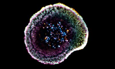
The researchers at Kyoto University observed this out-of-equilibrium behavior of wound healing during in vitro experiments with real cells on a substrate. They used a novel microscopy technique to allow them to measures protein activity within each cell: the protein was modified so that it lit up when activated thus revealing waves of protein activation propagating throughout the cell layer. The researchers were able to quantitatively predict the wave patterns, which they then also observed experimentally. More strikingly, they also found that the delay between the two waves was close to the theoretically predicted optimum for allowing cells to extract maximum information from the waves. This mechanism of self-organization is remarkable for allowing robust and spontaneous communication of direction over large distances within cell layers. It demonstrates one way in which coordinated behavior can arise in our bodies helping them to heal and grow.
Source: Institute of Science and Technology Austria (IST Austria)
30.09.2020