News • Potential treatment strategy
Vitiligo: pigment cells could be reactivated, study finds
Researchers find dormant pigment cells in vitiligo patches, suggesting a new treatment strategy

© oneinchpunch – stock.adobe.com
Vitiligo is an autoimmune disease in which the body's immune system attacks and destroys melanocytes, the cells that produce the skin pigment melanin. Once the melanocytes are gone, the skin turns white, leading to the characteristic skin patches of the disease. However, the standard model of vitiligo doesn’t explain some of its unusual features. Some vitiligo lesions re-pigment and treatments sometimes restore pigmentation even in areas that lack melanocytes, suggesting that the melanocytes may not be fully destroyed.
A team from Osaka Metropolitan University team led by Specially Appointed Professor Ichiro Katayama and Specially Appointed Associate Professor Lingli Yang has found evidence that melanocytes do not completely disappear from vitiligo skin and instead enter a “dedifferentiated-like state” in which the mature cells revert to a more primitive form, losing many of their specialized functions including the production of pigment. “This study uncovers a new mechanism underlying the development of vitiligo, which could change how we treat the disease,” Dr. Yang explained.
The findings were published in Nature Communications.
Their research adds to a body of research that shows that cells do not just respond to chemical signals, but also to what they are physically attached to. As melanocytes are located on the basement membrane, the thin layer that separates the epidermis from the dermis, the membrane provides instructions that help them remain functional pigment-producing cells.
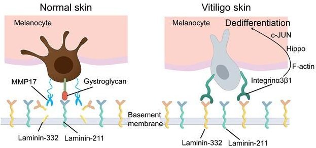
Image credit: Osaka Metropolitan University
However, the extracellular matrix surrounding melanocytes is remodeled in vitiligo patients. Usually melanocytes bond with laminin-211; however, in vitiligo patients, the basement membrane becomes enriched in laminin-332. Because their preferred binding partner is no longer available, melanocytes change how they attach. Instead of using their preferred attachment agent, dystroglycan, they attach through integrin α3β1 instead.
Most current treatments largely focus on suppressing autoimmune attacks and reducing inflammation, but if dormant melanocytes are still present in lesions that might change the way we treat the disease
Ichiro Katayama
These changes activate pathways that are commonly activated when cells are remodeling. In this process, the actin cytoskeleton of the cell is reorganized, gene expression is altered, including that of genes associated with an immature melanocyte state. The findings suggest a self-reinforcing cycle in which changes to the basement membrane drive melanocyte dedifferentiation, while the dedifferentiated melanocytes become less able to maintain a healthy basement membrane, further promoting the disease process.
“This was an exciting discovery for us, as most current treatments largely focus on suppressing autoimmune attacks and reducing inflammation, but if dormant melanocytes are still present in lesions that might change the way we treat the disease,” Professor Katayama said. “New treatment avenues such as reactivating existing cells or restoring their normal attachment to the basement membrane may be possible.”
When the researchers used pharmacological inhibitors targeting the signaling pathways activated by this adhesion switch, they were able to restore expression of mature melanocyte markers, recover pigmentation-related gene expression, and reverse many features of the dedifferentiated-like phenotype.
“This was an exciting aspect of our research, as it suggests that changing gene expression isn’t permanent and this process may be reversible,” Dr. Yang explained. “We found that drugs were able to restore melanocyte function and pigmentation-related characteristics. The next step will be to perform clinical studies to see if this approach is a viable way to manage the disease.”
Source: Osaka Metropolitan University
26.07.2026