Video: First whole-body MRI in Europe
Something that often obstructs a pioneering medical spirit is simply a practicality: the lack of space. For many hospitals, investment in new medical equipment is linked with construction and reshaping the hospital’s architecture – sometimes impossible because of the infrastructure. This was precisely the situation at the University Hospital Geneva (Hôpitaux Universitaires de Genève = HUG) when Professor Osman Ratib, Head of Nuclear Medicine, began to consider the installation of the first whole-body PET/MR to be installed in Europe.
Fortunately, he was only mildly disturbed by the space problem and far from forsaking the project just because of it. Consequently, the HUG and Philips Healthcare celebrated the inauguration of the innovative hybrid system in Geneva at the end of April – and they already presented some convincing results.
Visitors to the new site in Geneva generally experience two ‘Wow!’ effects. The first relates to the installation itself and the second to the way in which it was installed. The distinctive feature in the second is that the whole-body PET/MR was delivered on a truck – virtually ready to use: the system and all its components, such as injector room etc. was pre-installed in a container station 200 kms away from the hospital. This container was then transported to Geneva and placed directly against the hospital walls, and connected to the radiology department by a new door.
This door was the only hospital construction needed – and the only one possible due to the infrastructure. ‘For the installation of our new scanner we could benefit from a completely new design of a container that was conceptually devised by several small companies here in Switzerland,’ said Prof. Ratib. ‘The idea behind it was to pre-install the scanner in a large container that is specifically adapted to the size and the requirement of high technology equipment. This significantly reduced costs and time for the installation.’
From the February installation to the real premiere of a whole-body MRI took another three months. In April 2010 the first scan was performed and by the end of that month 25 patient examinations had been carried out.
Great potential for clinical use
So far, the combined scan by MRI and PET can only be perfomed in terms of clinical evaluations and research, because approval by FDA the is still missing. However, Prof. Ratib has predicted a huge potential for the future. ‘Today, many patients already have MRI and PET as part of their clinical follow-up and work-up, and get those two studies at two separate times, which means they have to come twice and have two different investigations done. We think it’s going to be a major improvement in patient comfort to be able to do the two studies at the same time in one single machine. ‘On the other hand, we believe that having those two modalities together will also improve the quality of our diagnoses and the accuracy of our diagnosis. We believe it will become a major player and have an impact in some of the new strategies and clinical work-up of some patients, especially in cancer and, more particularly, in evaluation of efficacy of some of these treatments and patient follow-ups to detect those who may, or may not, respond to the treatment being applied.’
Consequently, Prof. Ratib and his team will focus on evaluating the benefits of PET/MR for head and neck cancer, prostate cancer and breast cancer. The first examination results, which were shown during the inauguration presentation, were convincing. ‘MRI images provide anatomy and tissue characterisation combined with metabolic imaging from PET; we can now see tissue function and metabolism. In combination with new molecular tracer we are able to see and follow some tissue characterisation and activities
very precisly. So hybrid imaging is very promising, especially with regard to so called personalized medicine.
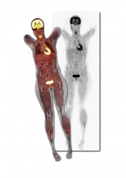
Whole-body PET/MR
The combination of the two modalities had long been a problem because the magnetic field created major artefacts during the PET scan. Philips Healthcare overcame this by providing sequential MRI and PET acquisition with stand-alone PET and MR scanners face-to-face, together with an innovative rotating bed, which accurately positions the patient inside each scanner. The installation in Geneva is the second worldwide, the other being at the Mount Sinai School of Medicine, New York. Both are equipped with a 3-Tesla MR and PET, using the latest Time of Flight (ToF) technology.
19.05.2010