Image source: Zhang R, Li L, Tang Y, Chen D, Journal of Intensive Medicine 2025 (CC BY-NC-ND 4.0)
News • Intensive medicine
Ultrasound-guided cannulation for safer vein access
A retrospective study evaluates the safety and efficacy of a novel ultrasound method for infraclavicular vein cannulation
Cannulation is a procedure of inserting a flexible tube (catheter) into the blood vessels to administer medication or to monitor hemodynamics. Inserting catheters into the major veins near the collarbone is associated with complications like pneumothorax. Therefore, accurate Cannulation without misplacement of the catheter is crucial. Ultrasound scanning techniques are being used to visualize the blood vessels for the correct placement of catheters. However, conventional methods do not provide a clear anatomical view for accurate Cannulation. A novel study presents a method called "Seeing Artery and VEin Simultaneously in the long axis" (SAVES) for ultrasound-guided infraclavicular axillary/subclavian vein cannulation, which demonstrated a 100% success rate without any serious complications.
This work was carried out by a team of researchers led by Dr. Lei Li and Dr. Dechang Chen, Department of Critical Care Medicine, Ruijin Hospital, Shanghai Jiao Tong University School of Medicine, China. Their work was published in the Journal of Intensive Medicine.
SAVES technique may be a safe and effective approach for ultrasound-guided infraclavicular axillary/subclavian vein cannulation
Lei Li
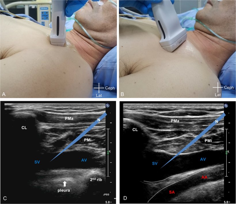
The SAVES method leverages the anatomical space created by the anterior scalene muscle between the subclavian vein and artery. Placing the ultrasound probe infraclavicularly provides a longitudinal view of both vessels' proximal (axillary) and distal (subclavian) parts while excluding the pleura from view. This approach offers theoretical advantages by potentially avoiding iatrogenic pneumothorax and reducing arterial damage risk. This study aimed to determine SAVES safety and efficacy for this procedure.
Researchers analyzed data from consecutive critically ill adults undergoing ultrasound-guided infraclavicular axillary/subclavian vein cannulation using SAVES by a single physician in a 12-bed medical/surgical ICU between August 20, 2021, and December 20, 2024. Outcomes measured included overall and first-pass success rates, access time, number of attempts, difficulties during insertion steps, and mechanical complications.
Results from 142 punctures across 111 patients showed:
- A 100% overall success rate.
- A 75.4% first-pass success rate.
- Median access time: 38.5 seconds (IQR: 21.5–80.0 s).
- Catheterization attempts: 1 attempt (88.7%), 2 attempts (9.2%), 3 attempts (2.1%).
- Guidewire insertion difficulty: 15.5% (No difficulties with dilator or catheter insertion).
- Hematoma formation: 2.1%.
- Posterior venous wall penetration: 3.5%.
- Catheter malposition: 5.6%.
- No instances of pneumothorax, hemothorax, arterial puncture, brachial plexus injury, or cardiac tamponade.
“SAVES technique may be a safe and effective approach for ultrasound-guided infraclavicular axillary/subclavian vein cannulation,” Dr. Lei Li stated. However, a larger controlled prospective study is warranted to confirm these findings.
In the classical approach, usually only one vessel (axillary/subclavian vein) is seen, and the pleura is in view, located beneath the vein. In the SAVES method, tilting the probe caudally allows visualization of the axillary/subclavian vein and artery simultaneously in the long axis, while the pleura is out of view. Theoretically, the SAVES method may avoid iatrogenic pneumothorax since the needle is pointed away rather than toward the pleura. The separation and space (the area outlined by the white dotted line) created by insertion of the anterior scalene muscle between the axillary/subclavian vein and artery. The pseudo-needle is almost parallel along the longitudinal axis of the axillary/subclavian artery, thus minimizing the risk of artery injury in the event of accidental penetration of the posterior wall of the subclavian vein.
Source: Journal of Intensive Medicine
14.09.2025