Ultrasound for very unusual patients
An opening lecture at WFUMB 2011 will surely excite the audience with its focus on using ultrasound examinations of larger, non-human patients. Delivered by Dr Thomas B Hildebrandt, head of the reproduction management research group at the Leibnis Institute for zoo & wildlife research in Berlin/Germany, delegates will hear of the ways in which veterinary needs push ultrasound to achieve intriguing and sometimes unexpected results. Report: David Ziska, ESR Office
Due to the unusual patients, ultrasound in veterinary medicine presents veterinarians and their equipment with a varied demands -- in particular, the corporality of some patients, whether major or minor, demands specific technical equipment as well as adjusted approaches, as Dr. Hildebrandt explains: ‘Sometimes, the size of some of our patients can be a real challenge. One extreme is scanning 1-2 mm ovaries of naked mole rates with an 80 MHz transducer; the other side is determining the position of baby elephants in the uterus with a 2 MHz transducer.
‘Another obstacle is that most of our patients need to be sedated before we can perform an ultrasound examination on them. Performing transrectal US on a male hyena with prostate problems without sedating it wouldn’t be a smart idea.’
Another task for Dr Hildebrandt and colleagues is facing the wide range of different species that are not entirely covered by the specialist literature. He spoke to us about, for example, his problems in identifying the position of the intra-abdominal testicles in Australian echidnas.
For veterinary ultrasound, in most cases there is a choice of only a limited range of equipment , but he counts himself a lucky exception: ‘Since the 80s, my research group has been in the lucky position to work with state-of-the-art ultrasound equipment. We use ultrasound microscopes up to 80MHz, 3-D/4-D high performance sonographs, portable 3-D/4-D ultrasound devices and the ultra-light brand new V-scan for missions in Africa or Asia’
Portable ultrasound devices are of great help in veterinary medicine but still struggle to provide high-resolution images and a high contrast level. To date there is no image of a 15-day-old bovine embryo.
To obtain such images portable ultrasound devices with a resolution up to 100 micrometers would be needed -- a resolution of which no mobile US device is currently capable. Looking to the future of portable US devices Dr Hildebrandt would greatly welcome the invention of wireless connections between monitor, processor and transducer, although he admits to the likely susceptibility of this to problems. Portable 30 MHz devices would also be very useful additions to the range of veterinary ultrasound devices.
Talking about the scientific hot topics at the WFUMB, he still foresees a long journey to reach the same level of ultrasound use as in human medicine: ‘In principle, classic veterinary medicine is about getting closer to the achievements of human medicine. So it’s very important to use events like the WFUMB 2011 to show to a broad audience what ultrasound in veterinary medicine is capable of. Sometimes even ultrasound experts in human medicine are quite surprised what we can achieve with ultrasound.’
Without revealing too many details about his talk at the WFUMB opening ceremony, Dr Hildebrandt agreed to give EH a little taster: ‘The cases I’ll present vary from ultrasonographic sex determination of octopuses and komodo dragons to gynaecological examinations of pachyderms such as elephants, hippopotami and rhinoceroses. In addition, I will recount a few things that have happened to us during our work with unusual patients.’
With his special fondness for Vienna, Dr Hildebrandt would also like to recommend Vienna’s zoo to the conference delegates: ‘At the ‘Tierpark Schönbrunn’, the world’s oldest zoo, our first test-tube elephant baby in Europe was born and we also did our first 3-D imaging of a baby elephant using a classic 530 Kretzmachine. Apart from being the oldest zoo the Tierpark Schönbrunn is also one of Europe’s role models for state-of-the-art animal keeping and I strongly recommend a visit while in Vienna.’
"Ultrasound in unusual patients", Friday, August 26, 18:00-18:20, Hall A
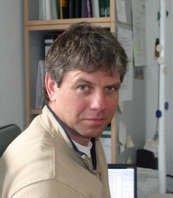
Thomas Hildebrandt
In 1987, veterinarian Dr Thomas Hildebrandt began his work at the Institute for Zoo Biology and Wildlife Research (ISW) in Berlin, and a decade later was appointed head of the institute’s new Reproduction Management Department, responsible for the development and establishment of the future direction of this research field.
The combination of basic and applied reproductive science in non-domestic species was responsible for the national and international success of his team seen in over 20 field projects as well as intensive collaborations with over 60 zoological institutions.
Dr Hildebrandt’s expertise in reproduction biology and pathology in elephants and rhinoceros is recognised worldwide. His development of a non-surgical AI technique in elephants was a key innovation that made artificial insemination successful in this species. It culminated in the successful insemination of six African and one Asian elephants over the last three years.
*Article originally published in the WFUMB e-Newsletter
25.08.2011