There is always urgency in pediatric radiology
Children will surprise you, and unfortunately they can sometimes hold big surprises for radiologists. At ECR 2010 four radiologists offered a series of pediatric cases in a session entitled, « Pediatric non-traumatic emergencies: What we must know!, » which covered children from head to toe, demonstrating that symptoms thought to be routine can mask more serious conditions
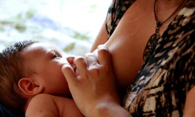
Dr. Catherine Garel, who specializes in pediatric radiology at the Debré Hospital in Paris, France, provided a compelling review of imaging for newborns that demonstrated the strengths and weaknesses of modalities encompassing ultrasound, computed tomgraphy (CT), magnetic resonance imaging and recent experiences with diffusion weighted imaging.
In one case, Dr. Garel said a child which presented with vomiting led the pediatrician to request adbominal examinations, but when no cause of the illness could be identified, she extended the investigation to include a brain scan, discovering damaged areas that were later related to child abuse.
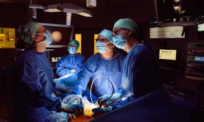
Dr. Charlotte de Lange, who specializes in thoracic imaging at the National Hospital in Oslo, Norway, told the audience that thoracic injuries and incidences are the most common non-traumatic urgent care cases among children.
“Always examine the airway,” she urged her colleagues when a child presents with breathing problems of difficulties swallowing as the small caliber of the trachea and its vunerability to collapse can quickly lead to more serious issues and that the smaller the child, the greater its vunerability.
She vividly made her point using CT angiographic scans, in one case of a child injured playing football whose greater injury was not related the bruising he received but turned out to be a stricture of his trachea.
Radiography remains the standard for examinations with ultrasound the first choice to more closely examine regions of interest.
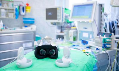
In pediatric emergencies, Dr. Simon Robben from the University Hospital Maastricht in the Netherlands, said most often it is not the crying child nor the anxious pareents that present as obstacle to an objective examination as the stressed-out pediatrician insisting on a specific diagnosis.
Remaining an detached specialist becomes essential for a radiologist to conduct a careful examination, as any delay to diagnosis increases the child's exposure to morbidity and, in some cases, mortality.
Following a radiography, also considered a standard for first examination, colon contrasted examinations are a necessary next step in the adomen where it is difficult, if not often impossible to distinguish the colon from the small bowel.
Ultrasound is the next choice for diagnostic validation, though not often conclusive, leading the radiologist who continues to have doubts or areas of concern toward the MRI scan , which he said is very useful to clearly see detail of the condtition or anatomical structure.
05.03.2010