The role of anaesthesia in MRI procedures
In response to increasing demand for anaesthesia procedures that complement MRI procedures, Dräger Medical is developing a new ventilator that will be compatible with field strengths of up to 3-Tesla
Currently, to expand and centralise the MRI Department at the University Hospital Schleswig-Holstein in Luebeck, Germany, a new building is being planned.
This will include the installation of two 1.5-Tesla MRI systems to cover most routine scans, and one 3-Tesla MRI system. The new MRI Department also plans to install Draeger Medical’s new MRI-compatible anaesthesia workstation, which includes all modern ventilation modes (volume and pressure controlled ventilation, SIMV/PS and PS), as well as compliance compensation. A new MRI-compatible patient monitor complements the workstation.
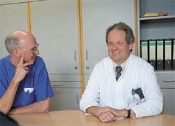
Professor Karl-Friedrich Klotz MD, Vice Director and head physician at the hospital’s Anaesthesiology Clinic, and Anaesthesia Nurse Stephan Hinz, who is responsible for medical practice guidelines, explained that, in the induction room, a standard anaesthesia machine will continue to be used. ‘The user interface of the older induction room anaesthesia machine cannot be compared with that of the new device,’ Prof Klotz pointed out, ‘but at present our personnel is familiar with it. In another step, a Fabius® Tiro could be added here, in order to simplify and unify the operating philosophy.’
Who would need anaesthetic during MRI scans?
High-acuity patients and, faced with confinement in a narrow MRI scanner, claustrophobic patients. Some of the latter, Stephan Hinz pointed out, only need reassurance and mild sedation (Medizolam), whilst others need more complex anaesthetic procedures, e.g. classic volume-controlled ventilation.
Sedation is also needed for neonates and older children treated by paediatricians, said Prof. Klotz. ‘For these we begin with Sevoflurane during induction and move on to intravenous or balanced anaesthesia. For the recovery phase we prefer Sevoflurane anaesthesia. Neonates and children should be ventilated during anaesthesia by pressure-controlled modes. Neonates weighing less than three kilograms require special attention and qualified equipment. The anaesthesia device currently installed in our MRI suite can only ventilate patients weighing more than five kilograms and does not have compliance compensation, which is advisable. And hand-bagging this very tiny patient through three metre-long hoses without proper compensation information is like flying blind. Hence, we do not usually anaesthetise neonates, but prepare them for MRI scans by sedating them.’
A large number of patients arriving from critical care wards also need anaesthesia in the MRI suite, Stephan Hinz added. ‘This group of high acuity patients is comprised of neurology, neurosurgery, and coma patients who require mandatory ventilation, as well as patients with blood clots, aneurysms, and epilepsy. These patients often arrive already connected to, or wearing, other devices, for example invasive blood-pressure measuring devices, pacers, infusions, Catecholamine therapy, and so on. Additional mandatory devices, such as syringe pumps, have to be arranged peripherally in the MRI suite; this procedure often includes elongating pump tubes. Easy measures, such as safety blankets, provide shielding for these additional devices.’
These intensive-care patients are often ventilated in quite sophisticated ways that must be modified during MRI scans, Prof Klotz pointed out. ‘Even pressure-controlled ventilation should be standardised from a therapy standpoint. We have to choose easier modes for device reasons. Intensive care patients are handed over to critical care ward personnel directly after the scan, to be transferred back to the ward.’
MRI patients need treatment at least once every other day, Stephan Hinz said, and sometimes scans can be scheduled three to four days in advance. However, then patients from critical care wards or trauma units often arrive unexpectedly, needing high staff input. ‘We have a special kind of logistical problem related to MRI scans,’ he said. ‘The MRI anaesthesia device is the only one of its kind in the university hospital. Therefore, not only are there special requirements and aspects of the delicate MRI environment that need attention, but also our staff is not routinely familiar with the user interface of the anaesthesia device. As said, many cases cannot be planned, so trained staff cannot be prescheduled. Therefore, we need to implement time-consuming training for our nurses and anaesthesiologists – over and over again – to have trained personnel available at all times.’
Prof Klotz estimated that, if obtaining an MR image takes six minutes, 30 minutes are needed for the anaesthetic procedure. But currently the MRI unit is in a distant part of the hospital campus, so the physicians and specialised nurses needed to prepared and support any unscheduled MRI procedure are taken away from their routine hospital work for up to four hours. ‘If you add the time needed to arrange all supplies and to prepare and check the anaesthesia device, it might easily consume ten hours of physician, nurse, and logistics personnel time. However, the hospital is only allowed to bill for the anaesthesia time, not the actual effort and time required.’
Presently, near the hospital’s MRI suite, the induction room is stocked with MRI compatible accessories, e.g. consumable patient hoses, soda lime canisters, MRI-specific ECG electrodes etc. Stephan Hinz explained. ‘MRI compatibility has to be checked by our anaesthesia nurses in advance. We use a non-MRI-compatible anaesthesia device and patient monitoring in the induction room.’
‘Whereas for adult patients, induction is mostly applied intravenously,’ said Prof Klotz , ‘we often use Sevoflurane for children to avoid venous in-dwelling cannulae, which might cause traumatic situations while the injection is taking place.’
‘The following steps comprise balanced and/or even intravenous anaesthesia,’ Stephan Hinz continued. ‘Leaving the induction room, the patient is placed directly on the MRI table, then connected to the MRI-compatible anaesthesia device, which has been checked and tested in advance. In our hospital, the MRI anaesthesia device stays in the MRI suite. Electrocardiograms (ECG) and partial, arterial oxygen saturation (SpO2) patient cables are specified for use in MRI environments.’
So, what would a ventilator need to offer to alleviate problems in the MRI suite – simply more compatibility with the imaging procedures?
The usual patient data is monitored (oxygen, ventilation pressure, tidal volume, carbon dioxide, and anaesthetic agent concentrations). ‘The most important ventilation modes are volume-controlled ventilation for healthy (even claustrophobic) patients and pressure-controlled ventilation for paediatric and critical care patients,’ Prof Klotz recapped. ‘So far, the more advanced ventilation modes, synchronised intermittent mandatory ventilation (SIMV) and pressure support (PS) for spontaneously breathing patients, have seldom been required. We have standardised our patient monitoring throughout the hospital. This is a tough task in the MRI suite because the MR environment requires various sensors, for example for ECG, non-invasive or invasive blood pressure (NIBP, IBP), and SpO2, and a dedicated user philosophy. To reduce the challenge to staff as much as possible, the differences between using a main operating room (OR) device and the MRI-compatible unit should be kept to a minimum. Unfortunately, this does not reflect the current status at our hospital.’
The ventilation and monitoring devices installed in the MRI suite are unique and require a high level of training and support to keep them up and running smoothly, Stephan Hinz pointed out.
Along with that, the anaesthesiologist is only needed if special patient conditions occur during the MRI procedure - for example, overcoming a 10–15 second apnoea phase, Prof Klotz explained. So, he added, mostly, the anaesthetist watches the MRI procedure through safety windows. Therefore, a larger display screen would be advantageous, he said, ‘…since the anaesthesiologist needs to get an angle on the vital curves and patient data provided by the anaesthesia workplace from outside. If the screens are too small for external viewing, an additional screen outside the MRI suite is needed, to display all important information. We currently use hand-written anaesthesia reports in the MRI suite. As soon as our hospital-wide automated record keeping system is installed, we will consider saving the MRI anaesthesia data directly in an electronic patient file.’
08.03.2007