The brain - A promising target for multimodal imaging
By Andreas Bauer MD, Professor of Functional and Molecular Neuro-imaging
Integrated PET/MRI systems will permit the simultaneous acquisition of molecular, functional and structural parameters. The combined strengths of PET (high sensitivity and specificity, but relatively low spatial resolution) and MRI (high resolution, but low sensitivity) is the most attractive feature of multimodal imaging with hybrid scanners. Their application could substantially contribute to the fields of basic as well as clinical brain research, because they deal with the most important problem of neuroscience: the relationship of structure and function.

Specific applications of integrated PET/MR systems for brain imaging are partly identical with applications for whole body scanners. For example, the identification of tumour tissue using amino acid tracers as [18F]-fluoroethyltyrosine ([18F]-FET) and [11C]-methionine is superior to stand-alone MRI. Amino acid PET improves the differential diagnosis particularly in cases of lymphoma, venous infarcts, dysembryoblastic tumours and recurrent tumours.
Although images of both modalities can be acquired by different scanners, and accordingly co-registered, a two-modality hybrid system will improve and simplify the fusion of biochemical and structural information. This holds particularly true if the fusion of imaging data is extended from structural to functional data e.g. to identify eloquent areas in the vicinity of tumours. Integrated PET/MR systems will be equipped with automated image fusion software and thus standardise and facilitate the co-registration of the PET and MRI data sets, which is primarily applied in research laboratories rather than a clinical environment, so far. Image fusion, for instance, has already become an important clinical tool in the early diagnosis of dementia and other neurodegenerative disorders, e.g., Huntington’s disease and multiple system atrophy. It is also important for the identification of structural changes in epileptic foci and can help to plan surgery.
A new dimension for brain research will undoubtedly derive from the fact that hybrid PET/MR systems not only enable combined but simultaneous data acquisition. During a given brain activation, it will be possible to identify the locally activated area with functional MRI (fMRI) while, in parallel, the underlying biochemical processes can be investigated with PET. Thus, multimodal imaging could substantially stimulate basic brain research. Furthermore, clinically oriented research will profit by the identification of molecular processes induced by local pathogenic factors as well as the consequences caused by innovative treatments, e.g. (stem) cell implantation and deep brain stimulation.
The identification of small regions or even cell clusters by MRI will thus become an essential prerequisite for the definition of ‘regions-of-interest’ to study biochemical processes by PET. Hybrid systems are therefore likely to stimulate the steadily growing field of invasive therapy approaches in neurological and psychiatric diseases.
PET/MR systems could also stimulate pharmacological imaging. Pharmacological MRI has become an important novel branch of functional MRI. In order to test the impact of a given drug, it is applied before or during fMRI. Local and regional changes of the blood-oxygen level dependent (BOLD) effect indicate drug-related alterations in information processing. To combine this type of study with molecular imaging would significantly increase the explanatory power of these investigations. PET would allow the study of the precise localisation and extent of receptor occupation or enzyme inhibition related to a given drug dose. This information is extremely useful because it can help to establish a definite link between the application of the drug and a given perfusional fMRI activation. Furthermore, PET allows the establishment of the pharmacodynamics and kinetics of the drug under investigation, which is a prerequisite to find an adequate dosage scheme.
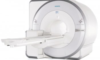
In 2009, an ultra-high-field PET/MRT system has been installed at the Jülich Research Centre in Germany, which will be used exclusively for brain research. Since resolution is critically related to field strength it is expected to push the resolution boundaries below the margin of 100 micrometers. This will allow study of the ctivity of single cell clusters. Real-time simultaneous studies will be of particular interest for the interpretation of activation patterns obtained with fMRI (vascular signal) and H215O PET (tissue signal). However, there are still significant problems to be solved. For instance, in technical terms we have to deal with the interaction of the magnet and the PET insert and its consequences for both modalities. An important physiological obstacle will be that the underlying processes of fMRI and PET have different time scales resulting in modality-specific differences in temporal resolution and sampling (time frames of seconds to minutes in fMRI versus acquisition times in the range of several minutes for metabolic and receptor PET).
In conclusion, integrated PET/MR systems hold great potentials for brain research, especially for multiparametric analyses of complex interactions of neuronal networks and their underlying brain chemistry. Multimodal imaging is also an interesting tool for the translation of new treatment strategies from preclinical research into clinical applications.
12.11.2009


