© PublicDomainPictures – pexels.com
News • Critical areas for osteoporotic fractures
Predicting hip fractures from just 7% of the bone
UPF scientists have discovered this 7% of critical areas based on their pioneering analysis model that allows minimizing the margin of error when predicting whether or not women with osteoporosis will suffer a hip fracture.
Scientists at Pompeu Fabra University (UPF) have made a great leap forward in predicting the risk of hip fracture among women due to osteoporosis. They have discovered that it is not necessary to examine all parts of the hip to predict whether it might fracture, rather it is sufficient to study 7% of the bone mass. It is the part formed by the so-called critical areas, and its identification is key to improving diagnostic methods, for example to narrow down the areas of the hip requiring an x-ray.
This will make it easier to design more personalized treatments that are tailored to their specific needs, for example, it will be possible to know which specific areas of the hip bone are most in need of calcium
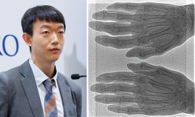
Simone Tassani
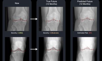
It has been possible to identify these critical areas thanks to the design of a new computational hip analysis model, which is unique in the world because it enables reducing the margin of error to 5% when predicting whether a woman will suffer a hip fracture due to osteoporosis. In other words, the degree of accuracy is 95% (according to the ROC-AUC classification method). This model is based on the 3D reconstruction of the hip using 2D x-rays and advanced statistical models.
In the study, the new analysis model was applied in a sample of 90 women with osteoporosis, 45 with a hip fracture and 45 without (control group). It is precisely by comparing 3D images of the hips of the two groups that enabled narrowing down the critical areas.
A recent article published in the journal Computers in Biology and Medicine has echoed the results of the research, led by Simone Tassani, a researcher at the BCN MedTech Research Unit of the UPF Department of Engineering. He has written the article together with Nicole Morando (main author), linked to the UPF research unit and to the Politecnico di Torino (Italy), and Carlos Ruiz Wills, a researcher at the BCN MedTech.
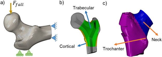
Image source: Morando N, Ruiz Wills C, Tassani S, Computers in Biology and Medicine 2025 (CC BY-NC-ND 4.0)
Osteoporotic hip fracture, the focus of this study, is growing worldwide due to the ageing population. Osteoporosis, which weakens the bones, especially affects the elderly and has a high incidence among women after menopause as the hormonal changes undergone at this stage in life hinder the regeneration of bone tissue.
This type of fracture can reduce patients’ mobility and quality of life and increase their risk of mortality. In the EU as a whole, the number of people with a fracture of the hip grew from 27.5 million to 32 million between 2010 and 2019. In Spain, there are some 50,000 cases of hip fracture per year, according to the National Register of Hip Fractures. In Catalonia, among major osteoporotic fractures (MOF), hip fracture is the most common and particularly affects older women. According to data for the period 2018-2020, hip MOF affects approximately 20 out of every 1000 women aged between 85 and 89 years; and about 12‰ between 80 and 84. In the case of men, these rates are close to 15 and 6‰, respectively (Government of Catalonia Ministry of Health, 2022).
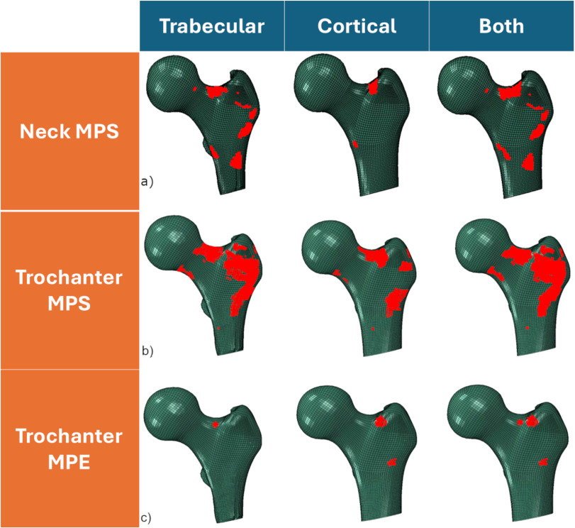
Image source: Morando N, Ruiz Wills C, Tassani S, Computers in Biology and Medicine 2025 (CC BY-NC-ND 4.0)
Thanks to the analysis model created by the UPF researchers, it will also be possible to better understand the specific causes of the process of bone tissue degeneration in each patient. “This will make it easier to design more personalized treatments that are tailored to their specific needs, for example, it will be possible to know which specific areas of the hip bone are most in need of calcium”, Simone Tassani (UPF) explains.
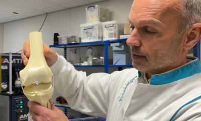
The new computational hip analysis model is based on previous methods created by UPF’s BCNMedTech Research Unit. These methods are based on a very common system used in engineering for calculating the impact that a movement or mechanical load has on the different parts of a structure (finite element model). Essentially, it consists of dividing the basic elements that make up a structure - based on its computerized three-dimensional representation - to calculate the impact of a virtual load or movement on each one.
Tassani clarifies that the application of this model on bones is especially complex: “In the case of bones, predicting the load caused by an impact or a movement is not as simple as with an object or a physical installation such as a bridge, because the materials of a bridge are uniform, but those of bones are not. In the case of a bridge, it is the point receiving the greatest load that will break, because everything is the same, but it not necessarily so in the case of bone tissue, whose mineral density, porosity, tissue orientation, etc. vary”. This study has enabled further adapting the previous model to the characteristics of hip bone tissue, complementing it with advanced statistical systems.
The scientists have applied the new analysis model to the 90 women participating in the study. They examined the differences in the critical areas of the hip of patients with and without fracture, not point by point but by grouping the areas under comparison, thanks to a complex statistical method (statistical parametric map), which reduces the margin of error to a minimum. The comparison of the two groups has allowed concluding that the risk of fracture is higher when the bone tissue of the critical areas of the hip is more uniform. Conversely, it is lower if the bone tissue is more varied.
The design of the new analysis model, the identification of critical areas and the results of comparative studies on women with and without hip fracture provide highly relevant conclusions, paving the way for improving the diagnosis and treatment of hip fracture among women.
Source: Pompeu Fabra University Barcelona
23.12.2025