Article • X-Nuclei MRI
Oxygen provides insights into tumour metabolism
Magnetic resonance imaging (MRI) usually measures the magnetic moment of the hydrogen atomic nuclei arising from the spin. However, scientists at the German Cancer Research Centre (DKFZ) are investigating the spin of other nuclei for imaging:
Report: Sascha Keutel
‘X-nuclei imaging has a large potential for MRI imaging as the x-nuclei play an important part in many physiological processes,’ according to doctor and physicist Daniel Paech MD, (Dipl. Phys). ‘For instance, we have been able to show that it is possible to measure oxygen-17 in the healthy brain and in brain tumour tissue and to gain information about the tumour’s metabolism.’
The researchers now published their findings in the journal Radiology.

The oxygen we breathe via air is mainly isotope oxygen-16. It has no magnetic moment, which is why it cannot be measured in an MRI scanner. The stable, non-radioactive oxygen-17 (17-O2) is also found but in smaller concentrations. To visualise the oxygen-dependent metabolism in the human body, such as for MRI examinations, or for dynamic inhalation experiments, the patient needs to inhale it in enriched form. Wherever oxygen is metabolised in the tissue, 17-O2 combines with hydrogen. This makes it detectable in the MRI scanner’s magnetic field. Tissue which metabolises a lot of oxygen shows up as lighter on the image. The radiologist would still be able to obtain an image without inhalation, but with poor resolution and without information on metabolic activity.
‘The exciting thing for us is that oxygen-17 can only be measured in its water-bound form, but not in haemoglobin-bound form. As oxygen is only consumed in one place in the body, i.e. at the mitochondrial membrane in the cells for energy production in the respiratory chain, we have a very specific method available to measure oxygen-dependent metabolism,’ Paech explains.
The Warburg effect
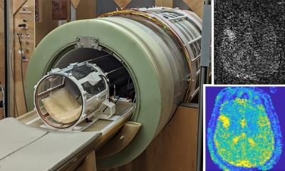
© Paech / Radiology
In the course of their technological development work, Paech and his colleagues initially tested the method on three healthy subjects. As expected, images of their brains showed a high metabolic rate (oxygen consumption). They then examined ten subjects with brain tumours. In these patients, lactic acid accumulated in the tumour cells. This metabolic product is the result of the anaerobic metabolism which the cancer cells prefer – even when they have sufficient oxygen available. This phenomenon is known as the Warburg effect. ‘With the help of oxygen x-nuclei imaging, we could show that the specific metabolism of the tumour can also be visualised in low-grade tumours, i.e. the tumour then shows as a region where no or hardly any oxygen is processed,’ Paech explains. To achieve a reasonable signal and good contrast, the researchers at the DKFZ developed specific coils for the 7-Tesla MRI which can currently only be used for MRI scans of the head.
Recommended article
Article • Maps of the brain
7-Tesla MR enters clinical routine
Ultra-high-field magnetic resonance tomography with field strength of 7 Tesla is slowly but surely entering clinical routine. ‘Thanks to very high spatial and spectral resolution, ultra-high-field MR permits detailed views of the human anatomy and can show precisely the metabolic processes such as those in the brain,’ said Professor Siegfried Trattnig, from Vienna’s Medical University.
Comparability
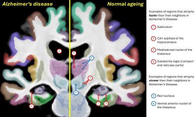
Normal magnetic resonance imaging is primarily structural – it visualises water in the body. Visualisation of tumour growth through changed structures and destroyed tissue is only a secondary effect. ‘The interesting feature of oxygen-17 imaging is that looking at the metabolic activity in the tissue takes things to a new level: It provides in vivo information about the tissue which should be viewed as complementary to the structural imaging techniques,’ the expert explains. Therefore, the researchers have not only gained new findings on the biology of brain tumours but also developed a new procedure that could help to characterise tumours more precisely, based on their particular metabolism. The researchers also expect that oxygen-MRI will provide valuable information for other diseases which go hand in hand with changes to metabolic processes such as Alzheimer’s disease or multiple sclerosis.
The only procedure currently directly comparable with 17-O2 MRI is oxygen-15-PET imaging. However, 15-O2 is radioactive and decays with a half-life of about two minutes. ‘We would require a facility for the production of 15-O2 right next to the hospital so that the isotope can be given to the patient within the shortest possible time. This is not practical in daily clinical routine,’ says Paech, in explaining why 15-O2-PET imaging has not become established.
Apart from hydrogen, there are further x-nuclei which could be of interest for medical imaging. However, as yet there are no clinical or scientifically relevant studies for potassium, chloride- or magnesium nuclei. Researchers have made some progress with natrium imaging which measures the natural distribution of natrium in the body. ‘Tumorous diseases result in changes to natrium signals as cell death,’ Paech reports, ‘and the development of oedema result in increased local natrium concentration.’
Profile:
Daniel Paech MD. Dipl. Phys. (2011) completed his medical studies in 2015 at the universities of Karlsruhe and Heidelberg and at the Sorbonne in Paris. He now heads the 7-Tesla MRI in Prof. Heinz-Peter Schlemmer’s department (E010 radiologist) at the German Cancer Research Centre in Heidelberg.
20.02.2020