New possibilities for the first trimester of pregnancy
New ultrasound technologies detect foetal heart defects earlier.
The Voluson E 8 includes several tools to help improve clinical workflow, such as Volume Computer Aided Diagnosis (VCAD), which considerably facilitates difficult heart examinations and delivers standardised images according to recommendations from leading obstetric and gynaecologic societies.
The system also provides a 4-D transvaginal probe, which gathers more precise data due to ultra-high frequencies and the new generation of probe technology.
At the congress of the International Society of Ultrasound in Obstetrics and Gynaecology, held in London this September), experts discussed the benefits of recent developments in ultrasound (US) technology, such as 4-D used in every-day diagnostic practice. During our interview with
Dr Greggory R DeVore, director of the Foetal Diagnostic Centre, in Pasadena, California,
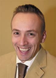
Dr Nick Raine-Fenning, of the Academic Division of Reproductive Medicine, University of Nottingham, where he is Consultant Gynaecologist & Clinical Senior Lecturer in Reproductive Medicine, and Professor Rabih Chaoui of the Department of Obstetrics and Gynaecology at the Charité, Berlin, it was clear that, in general, these advances are received with enthusiasm.
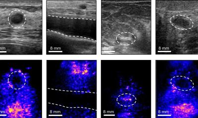
‘Thanks to this new ultrasound technology we can visualise, in the first trimester, details of the foetus that we have never seen before,’ said Professor Chaoui. For example, the high resolution of the new GE Healthcare ultrasound system produces images that enable examination of the foetal heart in the first trimester – when the foetus is just the size of a finger! In terms of size, Dr DeVore added that the US system is also ‘…small, mobile, easy to handle and provides images that could not be generated previously’.
Prior to this development, heart defects usually could not be diagnosed before week 22 of pregnancy. That progress is one effect of the 4-D technology, the experts agreed. Criticism that 4-D ultrasound is a nice tool for expectant parents, but not for diagnostic purposes, is unfounded, as Dr DeVore pointed out from his own experience with this system: ‘From a diagnostic point of view, 4-D is a real step forward, particularly for examining the foetal heart in week 7 or 10 of a pregnancy. With the new 4-D transvaginal probes, we can detect foetal abnormalities earlier than ever before, because they deliver volumetric information on even the smallest anatomic structures. Clinicians who are not specialised in cardiology can now more confidently evaluate the foetal heart, which may improve our overall prenatal detection rate of heart defects’
It is quite normal, says Nick Raine-Fenning, that some experts have a more critical opinion of
4-D ultrasound. After all, almost any medical innovation is initially smiled at. But there are always people, he adds, who recognise the potential of a new method, work with it, develop it and make it a success and thus, in the end, convince the sceptics.
One way to make prenatal exams of the heart more accessible for clinicians is to standardise diagnostic procedures. One of the new technologies is the so-called Volume Computer Aided Diagnosis, an automated imaging tool that makes volume imaging of foetal hearts less complicated by automatically generating multi-dimensional images of the right and left outflow tracts, once a standardised four-chamber view has been obtained. This technology provides clinicians with the potential to gather images that fit with the recommended standard screening views of a foetal heart published by the relevant societies.
‘Another step in this direction is “off line analysis”, because of the 3-D data storage. It allows manipulation of the dataset and images and gives the impression of actual patient examination. Clinicians can reassess the data at any time, using different viewing modalities to enhance the diagnostic ability,’ Nick Raine-Fenning added.
At this point, it is difficult to tell where ultrasound for obstetrics and gynaecology is heading. The new technologies generate data that have to be evaluated and interpreted. ‘We haven’t even grasped he potential of these new technologies. Quite obviously, if we can detect foetal heart diseases in the first weeks of pregnancy, the logical next step would be to treat the diseases as soon as we have diagnosed them. But this will take at least another 15 years,’ Professor Chaoui predicted.
Professor Rabih Chaoui MD, is a Board Member of the International Society of Obstetrics and Gynaecology, member of various German Societies, and on the editorial board of several national and international journals. He has authored and co-authored over 200 articles.
His special interest is cardiovascular haemodynamics in the foetus - foetal echocardiography and Doppler ultrasound. He has been using the Voluson since 2002.
2001 Prof. Obstetrics and Gynaecology in Berlin at the Charite Hospital
Since 2004 Private Centre for Prenatal Diagnosis and Human genetics in Berlin
Nick Raine-Fenning MRCOG MBChB PhD, is Associate Professor of Reproductive Medicine and Surgery at Nottingham University. Based at the Queen’s Medical Centre, he is involved with the University’s IVF unit (NURTURE) and the NHS Fertility Service. With a special interest in ultrasound, he recently established the Academic Imaging Suite, where his team carry out intensive research as well as provide a clinical service and active educational facility.
He was awarded a PhD for his thesis ‘The development and application of 3-D power Doppler angiography for the assessment of pelvic organ blood flow’. The majority of his work relates to infertility, but the unit has expanded interest to investigate endometriosis and dysmenorrhoea.
An expert in foetal ultrasound, obstetrician and gynaecologist Dr Greggory R DeVore has pioneered the identification of congenital heart defects using 2-D, 3-D, 4-D, and colour Doppler ultrasound. He has published numerous studies describing the use of foetal echocardiography to detect foetuses with Down’s Syndrome (in which, using genetic ultrasound, he has the highest detection rate reported in medical literature) and other chromosomal defects.
During medical training and as a Fellow in Maternal-Foetal Medicine at Yale University, he pioneered research on ultrasound evaluation of the foetal heart.
He became Associate Professor at the University of Southern California School of Medicine and is now a full-time consultant assisting community-based obstetricians with difficult foetal cases.
Dr DeVore has personally performed over 100,000 foetal ultrasound examinations. He has served on the editorial boards of medical journals, authored over 100 peer-reviewed papers, and contributed to more than 30 medical textbooks.
He also assists in the development of new ultrasound technologies for major multinational corporations.
17.11.2006