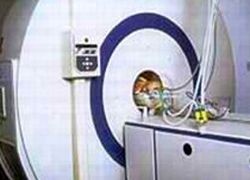
Neonatal MRI of premature babies
Infants born very prematurely have a high probability of experiencing behavioural and neuro-cognitive impairments during childhood and adolescence.
Globally, about 2% of all infants are born before 32 weeks of gestation. Due to medical advances in the last 20 years, around 85% of those infants survive in countries with hospitals equipped with neonatal intensive care units.
This impressive statistic is bittersweet, because the quality of life of very premature babies might be severely compromised. Almost 50% of these children will have significant cognitive, behavioural and social disabilities, and 10–15% will develop cerebral palsy and neurosensory impairment. The cost of years of medical treatment and special education is onerous for families, healthcare institutions, and the governments that pay for their citizens’ healthcare.
A global initiative by neonatal paediatric and imaging specialists has been underway since the mid-1990’s to utilize MRI imaging to identify brain abnormalities and injuries, so that treatments can be identified to minimize future disabilities. MRI is non-invasive and emits no radiation.
The Neonatal MRI Imaging Group at Imperial College London and Hammersmith Hospital, which have pioneered this initiative established the world’s first dedicated neonatal MR scanner for the neonatal ward in 1996, and garnered a second world’s ‘first’ with the only dedicated neonatal 3.0-T system, installed in 2005. The role of the Neonatal Neuroscience Research Group, headed by David Edwards MD, Professor in the Division of Clinical Sciences, Imperial College London, has been to understand the causes and consequences of neurodevelopmental impairment, so that strategies can be devised and initiated in the perinatal period of life. Numerous studies are in progress that utilize MRI imaging.
In Australia and New Zealand, at the Royal Women’s Hospital (Melbourne) and the Christchurch Women’s Hospital (Christchurch), a study conducted between 1998 and 2002 of 167 infants born at 30 weeks of gestation or less, strongly indicates that MRI brain scans become a mandatory standard of care for very premature infants. In the study, published in The New England Journal of Medicine (17/8/06), MRI brain scans performed at term equivalent could clearly display white-matter and grey-matter abnormalities.
To categorize infants according to the extent of their cerebral abnormalities, these were scored independently.
Of the 167 infants, 4% had severe, 17% had moderate, and 51% had mild white-matter abnormalities, and 49% had grey-matter abnormalities. 48 months later, after a comprehensive neurodevelopmental assessment of each child, the researchers found significant associations between the severity of identified abnormalities and the child’s subsequent risk of adverse neurological outcomes.
Research team member Terrie E Inder MD, Associate Professor of Paediatrics, Neurology and Radiology at Washington University School of Medicine (St. Louis, Missouri, USA) said the study results were important because MRI scans can predict the outcome of very premature infants better than any other clinical predictor, imaging predictor or combination thereof. She and her colleagues believe that the ability of MRI to detect changes that can be measured to monitor the impact of interventional treatment on the brain during the first 12–24 weeks of life in a neonatal ward will lead to discoveries of more effective treatments. She observed that similar results are being identified by the Neonatal MRI group at Hammersmith Hospital.
Utilization of medical technology that will improve an individual’s life is a priceless gift. Yet MRI imaging is expensive. Will the cost of routinely performing MRI scans of very premature infants, to identify those at greatest risk, lead to treatments that will reduce the overall medical and additional educational/societal cost of caring for these babies during their childhood and adolescence? The question is significant, and the answers are still to be determined.
14.11.2006