Nanomedicine
Inert inorganic particles reach selected tumour cells
Radiotherapy is a treatment of choice for six million (60%) of cancer patients annually. However, it has a crucial deficit: its therapeutic window is limited. The reason is simple. While radiotherapy is very efficient in killing cancer cells, it inevitably also damages surrounding healthy tissue. Therefore, in many cases doctors need to restrict the dose - a compromise that prevents the effective eradication of tumour cells.
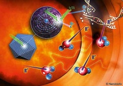
For decades, radiotherapists have not been able to greatly improve the situation. They believe that even a 5-20% increase in the therapeutic window could drastically change clinical output. French nanomedicine specialist Nanobiotix has developed NanoXray, a formulation of inert, inorganic particles with a size of 50nm that can be delivered selectively to tumour cells. Consisting of hafnium oxide, they react to a standard radiation dose by emitting a shower of electrons. This is the identical physical mode of action of radiotherapy – except that hafnium oxide emits far more electrons than a water molecule, the usual target, and thereby locally increases the effective dose by a factor of nine. The radiotherapy dose in the surrounding healthy tissue is unaffected because it is not loaded with nanoparticles.
NanoXray can be administered without the need to invest in novel devices; it does not even require changes in standard radiation procedures. NanoXray is applied either via intra-tumoural injection (NBTXR3) or by deposition of a gel-formulation in the tumour bed during surgical resection (NBTX-TOPO). A formulation for intravenous injection (NBTXIV) is also in development.
EU regulated as a medical device, NBTXR3 is in a Phase I study in soft tissue sarcoma patients. Preliminary results are expected at the end of 2012.
05.11.2012