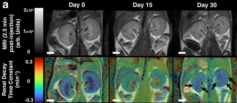
Image source: Calvert et al., Nature Communications 2023 (CC BY 4.0)
News • DCE-MRI urography
Metal-free kidney MRI dye for more personalized diagnostics
University of Ottawa scientists have introduced a metal-free MRI dye capable of mapping kidney function with unprecedented accuracy. This technique will modernize urology, allow for more personalized care and — ultimately — improve patient outcomes.
One in 10 Canadians suffer from some form of kidney disease. Current clinical methods for diagnosis rely on entering demographic and blood-based measurements into an equation to obtain an average value of kidney function for each patient. But this equation has a large error margin (around 20%) and doesn’t pinpoint which kidney is diseased or where the problem lies.
By proposing a new dye that addresses the safety limitations associated with traditional MRI contrast agents, this study opens the door to personalized diagnosis and treatment of kidney diseases. The dye can help obtain spatial data and quantitative glomerular filtration rate measurements for both kidneys, surpassing the limitations of current clinical methods.

Image source: University of Ottawa
“In speaking with radiologists and nephrologists, it became clear that we needed to do better. Magnetic resonance imaging (MRI) provides excellent anatomical images but lacks suitable contrast agents for reporting kidney function,” says Adam J. Shuhendler, associate professor in the Department of Chemistry and Biomolecular Sciences of the University of Ottawa’s Faculty of Science. “Existing MRI dyes with heavy metal atoms can be toxic if kidney function is impaired. This study addresses this limitation by introducing a metal-free MRI dye that enables safe and effective mapping of kidney function, providing crucial information for personalized diagnosis and treatment planning.”
The Canadian Institutes of Health Research-funded study, led by Nicholas D. Calvert, a PhD student in chemistry and biomolecular sciences in Shuhendler’s lab, was conducted with the help of Alexia Kirby, Mojmír Suchý, Peter Pallister, Aidan A. Torrens, Dylan Burger, Gerd Melkus and Nicola Schieda, from the Molecular Medicine Lab at the University of Ottawa and the preclinical MRI facility at the University of Ottawa Heart Institute.
Unlike the current equation-based method and traditional gadolinium-based MRI contrast agents, the novel dye enabled precise characterization of kidney function, even when one kidney was dysfunctional
Adam J. Shuhendler
The research team discovered a promising scaffold called verdazyl, which served as the basis for the metal-free MRI dye. After optimizing its synthesis, the dye was injected into healthy mice and imaged using MRI. The results demonstrated the efficacy and safety of the verdazyl dye, showing localization in the kidneys. Mouse models of kidney disease were subsequently used to validate the imaging-based method for mapping glomerular filtration rate (GFR). The results were compared with histology and established methods for determining GFR in mice, further confirming the reliability of the MRI-based approach.
“Unlike the current equation-based method and traditional gadolinium-based MRI contrast agents, the novel dye enabled precise characterization of kidney function, even when one kidney was dysfunctional,” says Shuhendler. “Remarkably, the study also revealed compensatory responses of healthy kidneys to their diseased counterparts.”
The introduction of the metal-free MRI dye represents a significant breakthrough in personalized urology. Using an accurate spatial map of kidney function, clinicians can develop tailored diagnosis and treatment plans for patients with kidney disease. The dye also presents as a chemical platform, which Shuhendler and his team are currently exploiting to target other organs and disease for MRI-based functional assessment. “With our new metal-free MRI dye, we have squarely entered the era of personalized medicine for kidney disease,” concludes Shuhendler.
Source: University of Ottawa
14.07.2023