News • Cancer research
Lymph node analysis to hunt down metastases
What makes tumor cells turn murderous? The Fraunhofer Institute for Toxicology and Experimental Medicine ITEM is investigating the mechanisms of metastasis formation – and searching for approaches for new treatments in the fight against cancer.
Among other things, the research team at Fraunhofer ITEM has developed a method that enables them to analyze entire lymph nodes.
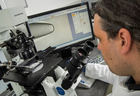
Single disseminated tumor cells can be fatal for cancer patients. They lie dormant in the bone marrow or other organs for years until, one day, they form metastases that ultimately lead to death. Nine out of ten cancer deaths are not caused by the primary but by secondary tumors. “We are developing special methods to genetically analyze single disseminated tumor cells and target them specifically,” explains Dr. Bernhard Polzer, deputy director of the Division of Personalized Tumor Therapy at Fraunhofer ITEM in Regensburg. The 40-member research team aims to use genetic data to shed light on the not yet well-understood process of metastasis formation and, in this way, to develop approaches for new forms of treatment.
With the conventional method, in contrast, pathologists found cancer cells in just 15 to 20 percent of the samples
Bernhard Polzer
Prof. Christoph Klein, director of the research division, sees great potential to avoid metastases in a diagnostic approach that learns to understand the onset of metastasis at the molecular and cell biological level. Important information for such an approach can be obtained by conducting a pathological examination of lymph nodes removed during surgical tumorectomy. For more than a hundred years now, when pathologists want to see whether a tumor has already disseminated, they search for cancer cells in ultra-thin slices of lymph node tissue taken from multiple locations. However, the tissue between these sections is disregarded, so any metastases located there remain undetected.
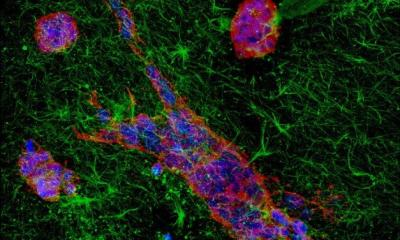
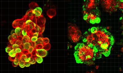
Together with the Fraunhofer Institute for Manufacturing Engineering and Automation IPA and the Fraunhofer Institute for Integrated Circuits IIS, Christoph Klein’s team has developed a method that makes it possible to analyze the entire lymph node. Samples are prepared using a tissue grinder, which carefully separates the cells. Afterwards, the sample is treated with a special dye that stains the disseminated tumor cells. Two million lymph node cells are scanned automatically and real cancer cells, as opposed to artifacts such as remnants of dye, are identified with high sensitivity by means of an AI-based imaging technique. “Using our method, we detected tumor cells in half of the examined lymph nodes,” says Bernhard Polzer. “With the conventional method, in contrast, pathologists found cancer cells in just 15 to 20 percent of the samples.” A market-ready version of the new method is currently being developed.
© Fraunhofer ITEM/P. Reinig
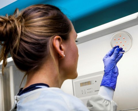
Once the single disseminated tumor cells have been isolated, their DNA must be replicated. Genetic analysis reveals, for instance, whether the disseminated tumor cells have the same properties as the primary tumor and might therefore respond to the same treatment. Often, however, they do not. The disseminated cells mutate when they reside in a foreign tissue environment for a longer period. “When breast cancer cells reside in bone marrow, they can take on the properties of the hematopoietic cells that surround them,” says Christoph Klein. “The cells evidently adapt to their surroundings.” But the disseminated cells can also activate additional cancer genes in their new environment. Then they become more and more aggressive and dangerous. If the genetic analysis reveals that the disseminated tumor cells have mutated, fighting them requires different remedies than those used for the primary tumor. Polzer and Klein are currently researching how to find the right drug for each patient.
Their starting point is a technology that already works for large numbers of cells and must now be adapted for single cells. It analyzes 450 cancer-specific mutations for which drugs are already available on the market or in clinical trials. Bernhard Polzer is clearly focused on the goal: “We want to isolate the disseminated tumor cells in lymph nodes or bone marrow and examine them for these 450 mutations. Then we could target them with the right drug to prevent metastasis.” To this end, the researchers in Regensburg are working with a number of companies that want to transfer this innovative diagnostic approach to everyday clinical routine.
Recommended article

Article • Cancer of unknown primary
CUP: in search for the smoking gun
Cancer of unknown primary (CUP) can send radiologists on a frustrating scavenger hunt: metastases were detected but the primary cancer is nowhere to be seen. Professor Alwin Krämer, Head of the Clinical Cooperation Unit Molecular Haematology/Oncology at University Hospital Heidelberg and the German Cancer Research Center, explains strategies for dealing with CUP.
When Bernhard Polzer carefully isolates a single disseminated tumor cell, he often asks himself: Would this cell have had the potential to kill the patient? Or would it have remained dormant and not done any harm? After all, not all disseminated cells form metastases. Christoph Klein calculated that, in a breast cancer patient with no visible metastases, there may be 10,000 to 20,000 disseminated cancer cells in the bone marrow. Only a few of those have the potential to metastasize. But which properties enable them to do this? Do they trick the immune system? Do they activate aggressive cancer genes? What turns them into killer cells?
To find an answer, Christoph Klein, in collaboration with several clinics, launched a study involving 200 breast cancer patients. His team examines these patients’ primary tumors, disseminated tumor cells and metastases. In each cell they examine, they analyze all the genes that cell activates. These data are then correlated with the patients’ clinical course – a vast amount of data to which the research group’s bioinformatics team gives structure. “We search the tumor cells for markers that are associated with a poor prognosis for the patient,” explains Christoph Klein. The full results of the study will be available by the end of the year – and will hopefully provide clues as to what turns a cell into a killer.
Source: Fraunhofer Institute for Toxicology and Experimental Medicine ITEM
03.06.2020


