Interview • Cardisiography
AI supported vector cardiography to enhance screening precision
Cardisiography is a precise screening method for detecting structural, rhythmical, or vascular heart disease. Neuronal networks assess and analyse vector cardiography data. The attending physician receives a report that describes a patient’s individual risk. Cardisiography is a decision support tool that guides the targeted follow-up for the patient. Meik Baumeister, CEO of Cardisio, gave insight into the development and the operating principle of the method.
Interview: Sara Hoffstedde
HiE: What is the idea behind Cardisiography?
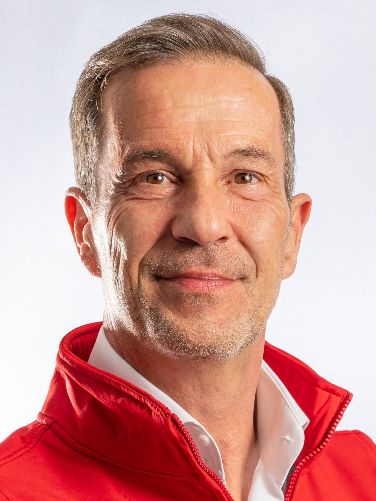
Image source: Cardisio
Baumeister: ‘In the summer of 2015, we had the idea to breathe new life into vector cardiography and find a way to apply it in clinical practice. The vector cardiography is a 70-year-old idea, with a lot of research on the technique showing great potential. Put simply, the method aims to map the heart and the propagation of the electrical excitation in multiple dimensions. However, interpretation of the multidimensional mapping of the heart is very complex. The mathematical analysis is barely possible with the human brain. Now, in the age of super computers and artificial intelligence (AI), we got a new chance to revive the dormant method using vector cardiography. Cardisiography unites neuronal networks with vector cardiography.’
How do vector cardiography and AI work together?
‘We measure normal medical parameters, such as the spacing or width of the QRS complexes. The two-dimensional ECG contains information about three angles. The three-dimensional VCG has information about four angles, a volume, and the sheathing surface of the measured loop. All these parameters are calculated and then analysed by the neuronal network. The calculation is followed by statistical evaluation, correlations are ruled out and significant results are identified. We use a big dataset to train the neural network in a supervised learning approach. Each patient is assigned their own set of parameters, which are compared to match the trained results for healthy or sick patients. This way, we can always reconstruct why the algorithm decided the way it did.’
What was crucial for the development?
‘Good data. We, as computer scientists, love code, math, and statistics, but without medical know how and good data we are lost. We needed time to get the data and to interpret the data successfully. We also needed – and still need – innovative cardiologists and open-minded medical doctors who declare their interest in the method and the willingness to use it. The cardiologists need to show that they trust the Cardisiography reports.’
How does Cardisiography work?
© German Cardiac Society
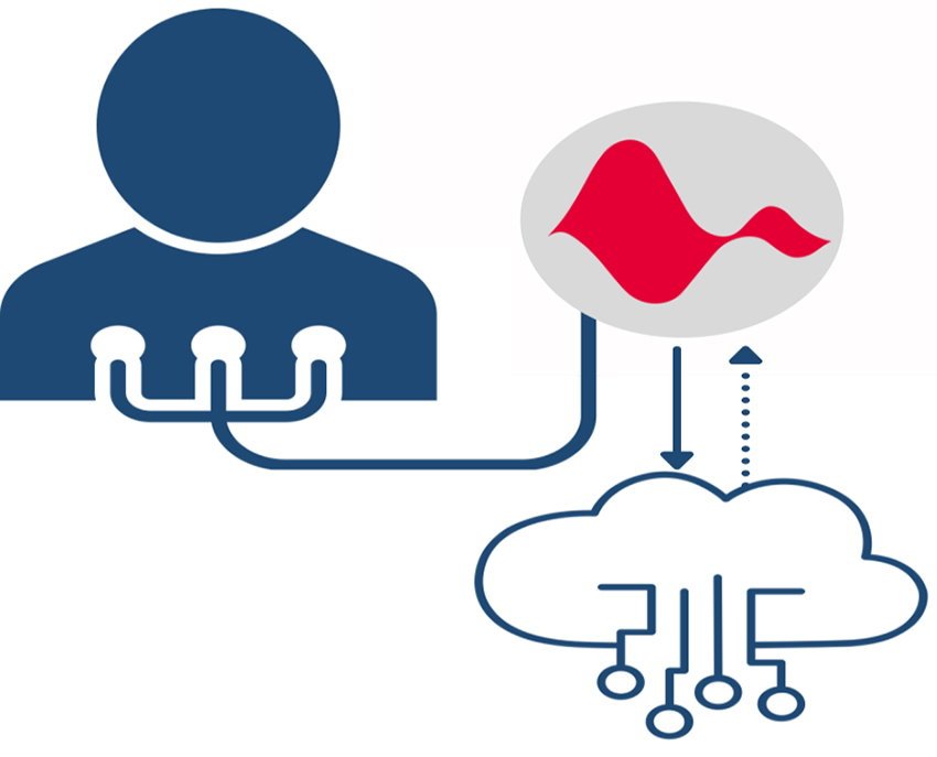
‘Three electrodes are applied to the front of a patient’s chest and another is added on the back. This establishes the “trigonum cardiacum” (heart triangle) and records the third dimension via the back electrode. While many argue that you can also get three-dimensional data with an ECG, that is just a projection. By using the back electrode, we get a native vector which is measured, not calculated. The hardware used to record the Cardisiography has to be adequate to compute all parameters measured. The recording itself is easy and only takes four minutes. After an additional four minutes, the results arrive. The recording does not need to be performed by a doctor; nurses or medical assistants can do it after being trained.’
How should the Cardisiography report be used?
‘The general practitioner, who used the Cardisiography, gets the report. He can read it and forward it to a cardiologist, if necessary. He can also hand it to the patient who can take it to other medical professionals. The report gives the specialist a pre-diagnosis, upon which a decision on further diagnostics and possible treatments can be made.’
Which information are included in the report?
‘Three areas are analysed: the perfusion of the heart, its structural integrity and rhythmology. Three bars (red to green) represent the patient’s risk for each of these areas. Information that is more detailed can be added for the expert, to serve as a basis for informed judgement. The additional data helps to identify the pathology of a given result, to exclude physiologically unusual cardiac positions as cause for that result.
‘If the algorithm finds a pattern, it is usually not just one parameter that indicates “not healthy”. Many patients with an inferior perfusion develop other heart problems. Because of the variable patterns, it is important that their roots are traceable. We use the three main areas to indicate where the algorithm found a pattern. The areas still include various diseases, but it gives the cardiologist a direction to look in.’
Which role do you see for the Cardisiography in the future of patient care?
With the method, the primary care physician is empowered to make a more qualified preliminary diagnosis and refer them to a cardiologist when they are at a serious risk
Meik Baumeister
‘Cardiovascular diseases are still the main cause of death. For people over 65, the prevalence is at 40%, with that number rising steeply with age. What this data does not show: people who get diagnosed at 65 started to develop the disease long before. Cardisiography is one part of the puzzle to optimize the prevention of cardiovascular disease. It is a screening tool that is very useful for preventive medical check-ups. If we were to decide, everyone from the age of 30 would be screened. Such an early screening could catch cardiovascular diseases while they are developing and before people go to see their doctor because of their symptoms.’
What is Cardisio's goal?
‘We want to send the right patients to the cardiologist. With the method, the primary care physician is empowered to make a more qualified preliminary diagnosis and refer them to a cardiologist when they are at a serious risk.’
Profile:
Meik Baumeister is co-founder and CEO of Cardisio. He has over 20 years of experience in implementing complex IT projects and leading IT companies. Starting as a consultant for business intelligence and CRM, his career as CEO led him to several medium-sized IT companies throughout Germany. Baumeister has particular expertise in the areas of e-health, cardiology and artificial intelligence.
26.08.2022