News • Combining common risk factors
Deep learning enables dual screening for cancer and CVD
Heart disease and cancer are the leading causes of death in the United States, and it’s increasingly understood that they share common risk factors, including tobacco use, diet, blood pressure, and obesity.
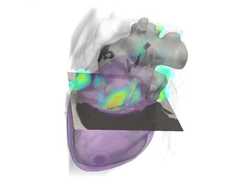
Image source: Chao et al., Nature Communications 2021 (CC BY 4.0)
Thus, a diagnostic tool that could screen for cardiovascular disease while a patient is already being screened for cancer has the potential to expedite a diagnosis, accelerate treatment, and improve patient outcomes.
In research published in Nature Communications, a team of engineers from Rensselaer Polytechnic Institute and clinicians from Massachusetts General Hospital developed a deep learning algorithm that can help assess a patient’s risk of cardiovascular disease with the same low-dose computerized tomography (CT) scan used to screen for lung cancer. This approach paves the way for more efficient, more cost-effective, and lower radiation diagnoses, without requiring patients to undergo a second CT scan. “In this paper, we demonstrate very good performance of a deep learning algorithm in identifying patients with cardiovascular diseases and predicting their mortality risks, which shows promise in converting lung cancer screening low-dose CT into a dual screening tool,” said Pingkun Yan, an assistant professor of biomedical engineering and member of the Center for Biotechnology and Interdisciplinary Studies (CBIS) at Rensselaer.
Numerous hurdles had to be overcome in order to make this dual screening possible. Low-dose CT images tend to have lower image quality and higher noise, making the features within an image harder to see. Using a large dataset from the National Lung Screening Trial (NLST), Yan and his team used data from more than 30,000 low-dose CT images to develop, train, and validate a deep learning algorithm capable of filtering out unwanted artifacts and noise, and extracting features needed for diagnosis. Researchers validated the algorithm using an additional 2,085 NLST images.
The Rensselaer team also partnered with Massachusetts General Hospital, where researchers were able to test this deep learning approach against state-of-the-art scans and the expertise of the hospital’s radiologists. The Rensselaer-developed algorithm, Yan said, not only proved to be highly effective in analyzing the risk of cardiovascular disease in high-risk patients using low-dose CT scans, but it also proved to be equally effective as radiologists in analyzing those images. In addition, the algorithm closely mimicked the performance of dedicated cardiac CT scans when it was tested on an independent dataset collected from 335 patients at Massachusetts General Hospital. “This innovative research is a prime example of the ways in which bioimaging and artificial intelligence can be combined to improve and deliver patient care with greater precision and safety,” said Deepak Vashishth, the director of CBIS.
Source: Rensselaer Polytechnic Institute
22.05.2021