Automated screening & reduced biopsies
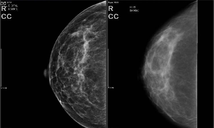
Although X-ray mammography can detect small cancers before they have spread. However, because abnormalities can only be identified non-specifically, percutaneous or surgical breast biopsy must follow - but less than 20% of women recalled for biopsies have cancer.
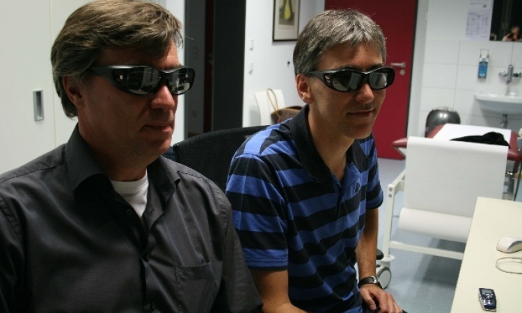
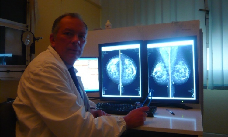
Now, research on the use of scattered X-rays has highlighted the potential for creating an automated process for breast cancer screening and reducing the need for biopsies. This work is among the many projects undertaken by the Synchrotron Radiation Department at the CLRC Daresbury Laboratory, UK, which span physics, chemistry, materials science, structural biology, engineering, environmental science, and novel applications to medicine and archaeology.
‘Invasive tumour expansion in breast carcinomas affects the collagen scaffold structure, a major component of breast tissue. Using small-angle X-ray scattering (SAXS), such changes in collagen structure are now detectable, and may lead to the characterisation of features in X-ray scatter distributions that show potential as disease markers,’ Daresbury Synchrotron explains. ‘If the molecular structure of the collagen is intact, the fraction of X-rays that pass through it appear in the form of peaks or rings, representing the effects of coherent interference caused by the diffracted rays. Peaks that are strong demonstrate healthy normal tissue, whereas peaks that are weak or diffuse indicate degraded tissue. The peak intensities have been shown to indicate conclusively which of the collagen specimens were cancerous and which were healthy.’
Preliminary results suggest that this technique can be used to make accurate assessments of cancerous versus normal breast tissue, and also for the detection of benign tumours,’ the lab points out. ‘There is also scope for in vivo application, which would both eliminate the need for breast tissue removal and greatly reduce the analysis time compared to that of current methods.’
Details: www.srs.dl.ac.uk
01.07.2004