Giotto - The Italian route towards digital breast tomosynthesis
It’s digital mammography taken to the next level – or, so to speak, the next dimension as digital breast tomosynthesis (DBT) that provides high resolution 3-D imaging. For about two years this exciting technology has promised to become the magic bullet in the early detection of breast cancer, particularly in the dense breast.

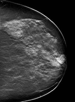
However, following initial euphoria, things quietened down about DBT -- mainly because the mandatory quality control guidelines and FDA approval for the US market are lacking. Italian firm I.M.S. is now working flat out to produce a patented optimisation of a second generation DBT to their Giotto Image 3-D mammography system.
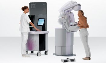
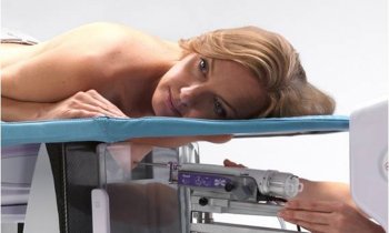
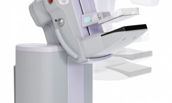
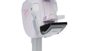
I.M.S. based in Bologna is mostly known for the ring-shaped gantry design of their Giotto mammography unit. In 2003, the firm was the first to introduce Full Field Digital Mammography (FFDM) in Europe. A year later, they were the first, worldwide, to use FFDM for stereotactic biopsy examinations. Indeed, I.M.S. can really be named as a pioneer in breast imaging technology. Now, they are conducting the largest multicentre clinical trial on DBT technology. Involved are the university radiology departments of Bologna, Milano, Udine, Stockholm and Maastricht.
Starting in November 2010, 1,010 screening patients with dense breast and 50 patients with suspicious findings are being examined with the new DBT system, with first results due to be published six months later. ‘We truly want to validate the utility of our system before entering the sales market,’ explained Achille Albanese, Marketing Director at IMS, during our European Hospital interview. ‘The IMS DBT system now being tested by our clinical partners is the outcome of ten years of R&D into advanced reconstruction methods, optimal imaging geometries, variable dose distributions and novel tomosynthesis visualisation methods.’
DBT means low dose, limited angle X-ray tomography of the breast. While in FFDM the X-ray source acquires 2-D projection images statically from only one point, in DBT the tubes move in an arc around the breast. The 2-D projection images are then reconstructed on a computer to form a 3-D volume made up of 1mm slices through the breast. Thus, while in FFDM, lesions can be overlapped by breast tissue and remain undetected, DBT ‘peels away’ these layers of tissue.
So, what is the difference in the IMS DBT technique? ‘In contrast to other available tomosynthesis systems on the market we depart from CT style, filtered back projection reconstruction algorithms and geometries, with uniform distribution of dose and projection angles,’ Achille Albanese emphasised. ‘We rely instead on variable choice of angles and dose. The biggest limitation in tomosynthesis is, of course, that it should not exceed the dose of a normal mammogram. At the same time, we certainly want to acquire a high image quality that can provide more clinical information than FFDM. There is a complex of interrelated factors affecting image results in tomosynthesis. One is the angular range: While a wide range gives better depth information, a narrow range is quicker in examination. So we choose to a combination of both, which is a width of 40°.
‘Another important factor is number of exposures,’ he added. ‘Fewer projections improve the signal to noise ratio in each projection. But while other manufacturers prescribe a certain exposure, we decided on a choice of dose, so that the radiologist can decide in which areas he needs more or less exposure.’ And, he pointed out, ‘Another specialty of this optimised variable dose geometry is that it enables the radiologist to use sufficient dose in the central projection to be a 2-D mammogram. So instead of increasing the X-ray ‘dose budget’ by first doing tomosynthesis and subsequently mammography, the reconstruction algorithm generates both image data into one 3-D image.’
Among those currently working with the Giotto system is radiologist Dr Gianni Saguatti, Head of the Breast Care Department at Ospedale Maggiore, Bologna, Italy, who said: ‘My experience concerning the use of the Digital Breast Tomosynthesis gives me the opportunity to emphasise the top features this method has shown. The opportunity to carry out a serial study of the mammary volume allows us to focus accurately on lesions, their morphological aspects as well as their real radiological size. Our Centre is now going to start an ad hoc clinical trial to test this application in the mammographic screening survey and in the perspective of a better detection of cases of multifocality.’
As soon as IMS DBT technology shows adequate results in the European clinical study it will be integrated into the Giotto Image 3-D. Achille Albanese believes this could be by mid-2011.
He also revealed that tomosynthesis is only one of the advanced applications currently a work-in-progress at I.M.S. ‘The high power X-ray generator of 8 kW we use for tomosynthesis is also prearranged for other new techniques, such as dual energy and, in this context, contrast media mammography.’
Dual energy generates two images with two exposures: One with low energy for fatty tissue and one with high energy for glandular tissue. This enables the subtraction of fatty and glandular tissue of the breast image, so that only abnormalities are shown. ‘What we still need is the implementation of a new X-ray sequence,’ he predicted. ‘This will be our next R&D project, as soon as we are successfully finalised with tomosynthesis.’
02.03.2011
- breast cancer (625)
- breast imaging (125)
- imaging (1639)
- mammography (258)
- medical technology (1554)
- women's health (337)
- X-ray (297)


