Artificial blood vessels can grow with the recipient
In a groundbreaking new study led by University of Minnesota biomedical engineers, artificial blood vessels bioengineered in the lab and implanted in young lambs are capable of growth within the recipient. If confirmed in humans, these new vessel grafts would prevent the need for repeated surgeries in some children with congenital heart defects.

One of the greatest challenges in vessel bioengineering is designing a vessel that will grow with its new owner. In this study, University of Minnesota Department of Biomedical Engineering Professor Robert Tranquillo and his colleagues generated vessel-like tubes in the lab from a post-natal donor’s skin cells and then removed the cells to minimize the chance of rejection. This also means the vessels can be stored and implanted when they are needed, without the need for customized cell growth of the recipient. When implanted in a lamb, the tube was then repopulated by the recipient’s own cells allowing it to grow.
“This might be the first time we have an ‘off-the-shelf’ material that doctors can implant in a patient, and it can grow in the body,” Tranquillo said. “In the future, this could potentially mean one surgery instead of five or more surgeries that some children with heart defects have before adulthood.”
To develop the material for this study, researchers combined sheep skin cells in a gelatin-like material, called fibrin, in the form of a tube and then rhythmically pumped in nutrients necessary for cell growth using a bioreactor for up to five weeks. The pumping bioreactor provided both nutrients and “exercise” to strengthen and stiffen the tube. The bioreactor, developed with Zeeshan Syedain, a senior research associate in Tranquillo’s lab, was a key component of developing the bioartificial vessel to be stronger than a native artery so it wouldn’t burst in the patient.
The researchers then used special detergents to wash away all the sheep cells, leaving behind a cell-free matrix that does not cause immune reaction when implanted. When the vessel graft replaced a part of the pulmonary artery in three lambs at five weeks of age, the implanted vessels were soon populated by the lambs’ own cells, causing the vessel to bend its shape and grow together with the recipient until adulthood.
“What’s important is that when the graft was implanted in the sheep, the cells repopulated the blood vessel tube matrix,” Tranquillo said. “If the cells don’t repopulate the graft, the vessel can’t grow. This is the perfect marriage between tissue engineering and regenerative medicine where tissue is grown in the lab and then, after implanting the decellularized tissue, the natural processes of the recipient’s body makes it a living tissue again.”
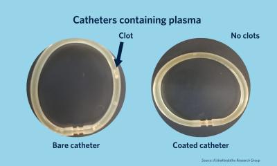
At 50 weeks of age, the sheep’s blood vessel graft had increased 56 percent in diameter and the amount of blood that could be pumped through the vessel increased 216 percent. The collagen protein also had increased 465 percent, proving that the vessel had not merely stretched but had actually grown. No adverse effects such as clotting, vessel narrowing, or calcification were observed. “We saw growth and none of the bad things happened,” Tranquillo said. “The results are very encouraging.”
Tranquillo said the next step is talking with doctors to determine the feasibility of requesting approval from the Food and Drug Administration (FDA) for human clinical trials within the next few years.
Source: University of Minnesota College of Science and Engineering
06.10.2016


