News • Women's health
Adenomyosis: New insights on under-recognised condition
Researchers at the University of Liverpool have identified distinctive biological characteristics within adenomyosis lesions that could help pave the way for more targeted, less invasive treatments.

© nenetus – stock.adobe.com
The findings from a team based in Professor Dharani Hapangama’s gynaecology research group at the University, provide new insights into the common but often under-recognised condition that affects up to one in five women of reproductive age. Despite its prevalence and significant impact on health and quality of life, adenomyosis has historically received limited attention in research and clinical practice.
The paper was published in the journal Science Advances.
Adenomyosis occurs when tissue similar to the lining of the womb grows deep into the muscle of the womb. It can cause heavy menstrual bleeding, severe pain, and, in some cases, pregnancy and fertility complications. Once thought to mainly affect older women, adenomyosis is now recognised as being common among younger women, including those who may wish to have children. Growing awareness of the condition has highlighted the need for improved diagnosis and more effective treatment options. Current treatment options remain limited, largely consisting of hormone-based therapies that can prevent pregnancy or surgery to remove the womb. The lack of treatment choices reflects longstanding gaps in understanding the biological mechanisms that drive the disease.
The new study sought to understand how diseased tissue differs from healthy tissue within the uterus. By identifying the unique biological features of adenomyosis lesions, the research aims to support the development of treatments that target affected areas while preserving healthy uterine tissue.
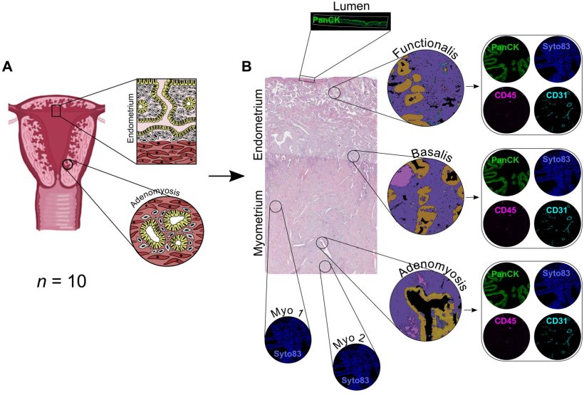
Image source: Maclean A, Johnson E, Rushworth C et al., Science Advances 2026 (CC BY 4.0)
To investigate these differences, researchers used spatial transcriptomics, an advanced technique that enables scientists to examine which genes are active in different cell types within their original tissue environment. Used for the first time to study human uterine tissue, this technique enabled direct comparisons between diseased and healthy tissue from the same uterus while preserving the tissue’s structural integrity – a capability that previous methods lacked.
By identifying the features that distinguish diseased tissue from healthy tissue, we are laying the groundwork for treatments that could directly target the condition while preserving the uterus and normal womb lining
Alison Maclean
The findings revealed that adenomyosis lesions possess a distinct biological “fingerprint”, suggesting the possibility of developing treatments that specifically target lesions while leaving healthy tissue intact. Researchers also found that the lesions share characteristics with the deeper, more stable layer of the womb lining, which may help explain how the condition develops and persists over time.
In addition, the study identified evidence of ongoing inflammation and altered energy production within the diseased tissue. These processes may contribute to some of the symptoms commonly experienced by women with adenomyosis, including pain and heavy bleeding.
Using the molecular information generated by the study, researchers also identified a number of existing drugs and emerging medicines that could potentially reverse some of the biological changes observed in the lesions. While further investigation is needed, these findings may help guide the development of future treatment options that do not require major surgery.
Dr Alison Maclean, NIHR Clinical Lecturer, who undertook this study during her MRC clinical research training fellowship said: “This study provides important new insights into what is happening within adenomyosis lesions at a molecular level. By identifying the features that distinguish diseased tissue from healthy tissue, we are laying the groundwork for treatments that could directly target the condition while preserving the uterus and normal womb lining. Ultimately, our goal is to develop safe and effective therapies that help women avoid major surgery and protect their fertility where possible.”
Source: University of Liverpool
08.07.2026