Image source: TU/e; photo: Bart van Overbeeke Fotografie
News • 'Sweet' assistance
3D printed vascular models to help cancer therapy
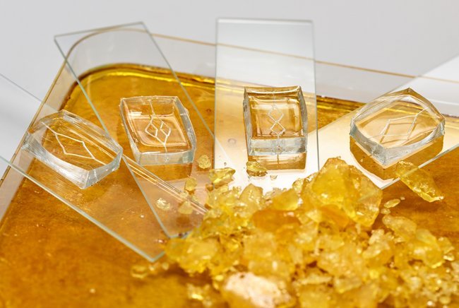
3D-printed sugar models of dense and chaotic blood vessel networks near tumors could help future cancer treatments.
To fight cancer, imaging, treatment, and monitoring are all needed. In contrast to blood vessels in healthy tissue, a denser and chaotic blood vessel network is an indicator of a tumor. A promising new way to study blood flow in these vessels involves injecting air bubbles and then tracking them with ultrasound. This shows where the blood goes and how the vessels are working, as well as helping with diagnosis, but it can be hard to see bubbles in ultrasound images of real blood vessels. As a ‘sweet’ alternative, PhD researcher Andreas Pollet from the Eindhoven University of Technology (TU/e) turned to sugar to make 3D-printed mock-ups of blood vessel networks to more easily study blood flow near tumors.
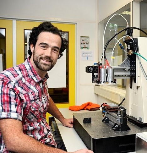
Image source: TU/e; photo: Bart van Overbeeke Fotografie
According to Mary Poppins “A spoonful of sugar helps the medicine go down”, but PhD researcher Andreas Pollet would argue that sugar could do a lot more than that in healthcare. “Sugars are important for a whole host of bodily functions. But in the lab, sugars can be used to build complex models of systems in the human body such as blood vessel networks,” says Pollet, who completed his research in the group of Jaap den Toonder in the department of Mechanical Engineering. “And key to the whole process is 3D printing.”
3D printing is ubiquitous in society today. With the right materials, you can 3D print almost anything from concrete homes to chess pieces and chocolate desserts. For those with a sweet tooth, it might be disappointing to learn that Pollet was not aiming to 3D print tasty sweet desserts. Along with his collaborators, Pollet designed a custom 3D printer that can very precisely print fibers of sugar. Multiple fibers can then be combined to create networks that look like blood vessel networks. “Sugar allows for printing without rigid supports, and the material changes from a liquid to a solid after cooling. It can support its own weight then. Furthermore, it’s biocompatible and dissolves in water – something not normally associated with 3D printing materials,” says Pollet.
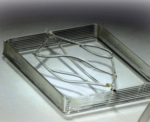
Image source: TU/e; photo: Andreas Pollet
Once the blood vessel model is printed, it’s time to say goodbye to the sugar. Pollet: “The sugar is just used to make a mold of a blood vessel network. We can’t use a model of a blood vessel network made of sugar to study blood flow as it would dissolve straight away. So, we coat the network, and then cast it with another material.” The sugary networks are coated with a biocompatible and biodegradable polymer to prevent unplanned dissolving of the sugar during casting. Once coated, the networks are cast using PDMS, a widely used silicon-based organic polymer that gives long-term stability, or hydrogels, which have properties like human tissue and can be easily imaged with ultrasound.
Pollet’s use of sugar to make the blood vessel networks can be hugely beneficial when it comes to making artificial blood vessels, particularly small blood vessels. “We can print vessels with diameters from several millimeters to one tenth of a millimeter (same thickness of a hair). And we can print branching points in the vessels, just as you would expect to see in blood vessel networks in the body,” says Pollet. “The control is phenomenal.”
Motion of air bubbles or fluids through the printed networks can be tracked using ultrasound imaging and microscopy to provide information on blood flows through the networks, with Pollet’s research colleagues Peiran Chen and Meiyi Zhou working on signal processing and ultrasound hardware development respectively. Results from the imaging approaches can then be compared.
Additionally, to make the vessels in the networks that work like real blood vessels, Pollet lined the inside of the network channels with cells from blood vessels. “To keep the cells alive, we placed cell culture medium in the vessels and supplied them with fluids to survive.” Besides 3D sugar printing, Pollet also used two other ways to build blood vessel networks. First, he used columns of different sized beads to approximate vessel networks where columns of larger beads are healthy networks, while columns of smaller beads are networks around tumors. In the second approach, Pollet grew intricate blood vessel networks using real cells in a growth chamber.
The next step for Pollet is a postdoctoral position at Sanquin in Amsterdam, who are responsible for blood supply in the Netherlands on a not-for-profit basis, in a project together with TU/e. “The goal is to mimic bone marrow. We want to devise a way to make platelets and red blood cells on a chip. Like my PhD research, the project is linked to the vasculature and blood flow.”
Pollet's research has shown that you need a range of materials to make blood vessel networks in the lab. Real cells, polymers, and hydrogels all play their part. But it’s the use of sugar that seems the most unconventional. Yet as it turns out, it’s perhaps the most effective and important.
Even Mary Poppins would be impressed.
Source: Eindhoven University of Technology
02.04.2022


