Article • An era of turbulence and innovation
The birth and rebirth of imaging
The New Horizons Lecture at the RSNA annual meeting is a keynote address that looks to the future, and the inventor of a major innovation in magnetic resonance imaging (MRI) technology, Daniel K Sodickson MD PhD, did just that. His lecture entitled ‘A New Light: The Birth and Rebirth of Imaging’ looked back at how MRI has evolved and forward at what it will become.
Report: Cynthia Keen
The change will be radical. Today’s radiology images may be irrelevant. MR protocols will become obsolete – but radiologists will not be, Sodickson emphasised, because radiologists are in a position to transform the field. ‘We invented diagnostic imaging. We figured out how to visualise and understand human inner space. This is both a turbulent age for imaging and a golden age for innovation. We need to embrace the theme of RSNA 2017, and ‘Explore, Invent, Transform.’ Modern imaging science is information science. Let us be the scientists who create new forms of information. In a world increasingly dominated by information, what more valuable contribution can we make?’
Sodickson explained that the era of continuous, comprehensive imaging data has arrived, and that with this change, the era of the ‘carefully framed snapshot’ of a high-resolution image will pass. ‘Radiology is not just copying our eyes. Instead, it is starting to emulate the way that our brains process multiple, streaming multi-sensory experiences.’ In his lecture Sodickson described the building blocks that created MR technology, from 1973 when the idea was first published to the revolutionary changes and relentless innovation that have ensued. This rapid-fire overview clearly illustrated how radiology progresses, adapts, and advances diagnoses through imaging.
Dramatic improvements of the 1990s

Sodickson spoke of the evolution from the first magnetic field gradients to diffusion imaging. The introduction of functional MRI, implemented in 1990, enabled radiologists to see different types of biophysics, reflecting brain activity. In the 1990s, some dramatic improvements were made to gradients and to radiofrequency coils, enabling advances in speed and image information content. The concept of moving from a sequential imaging device to a parallel acquisition device decreased the time to acquire images. Parallel acquisition made it possible to reduce corruption of images by motion from the abdomen and heart, and also to make the process easier for patients.
In 2007, compressed sensing was introduced. In addition to making the acquisition of images faster, it made feasible the use of multiple dimensions of data, which could be integrated together. Non-traditional data sampling patterns, such as radial patterns, gained new prominence. One particular example Sodickson described used a golden angle radial pattern, which enabled motion robustness during continuous data acquisition.
With the introduction of continuous three-dimensional acquisition, it became possible to perform multidimensional sorting along distinct motion dimensions. This enabled the creation of unique cardiac motion dimensions or respiratory motion dimensions, as an example. More innovations enabled radiologists to determine how the heart contracts and relaxes at any stage, or to look at the morphology of the great vessels or coronary arteries and freeze the images at any stage, thus enabling better heart disease diagnoses.
Creating a dictionary of pre-simulated fingerprints
The addition of 4-D, 5-D, and 6-D image processing algorithms led radiologists away from a mode of carefully tailored and adjusted snapshots toward a much simpler paradigm of rapid and continuous streaming images. A variant of this paradigm, magnetic resonance fingerprinting, was developed in 2013. Rather than standard image reconstruction, MR fingerprinting created a dictionary of pre-simulated fingerprints for different types of tissue and matched acquired singles to these fingerprints to create quantitative maps of tissue properties. MR fingerprinting offers the promise of scanner- and operator-independent scanning. If this promise is delivered, the feasibility of conducting clinical trials in radiology with tens of patients could easily expand to thousands, thus making clinical trials more accurate. Findings of such huge clinical trials could advance precision radiology.
Assessing a continuously acquired multidimensional data stream
If we stay true to our rich history of invention, we are bound to see things in a new light
Daniel Sodickson
Enter artificial intelligence (AI). ‘The artificial intelligence I see benefiting radiology is not merely image interpretation but rather data interpretation,’ Sodickson said. ‘AI neural networks can learn the various tricks of parallel imaging, compressed sensing, and other transforms we don’t know yet. ‘These could create images that are better than ones any existing image processing algorithms can produce and by doing so, they can help radiologists to make better diagnoses.’’ This is happening now, he said; but is happening with single slices and static images. What if you could have an AI neural network to assess a continuously acquired multidimensional data stream? AI would be the perfect way to economically produce actionable information, with biological fingerprints of disease as the output.
However, if AI could determine all this, would images even be needed? And how would MR scanners change? Could MR scanners be stripped down to the bare minimum of simply acquiring continuous data with the push of a button? And what would be the role of radiologists? Sodickson has suggested that, in the new era radiologists are facing, they would be information traffic controllers. They need to control and interpret the information content and context, and make it relevant for the treatment of patients. ‘We too are a force for change, a juggernaut of extended vision and expanded mind. If we embrace emerging paradigms of technology and information, and put them to good use, we will continue to see what was once invisible. If we stay true to our rich history of invention,’ he concluded, ‘we are bound to see things in a new light.’
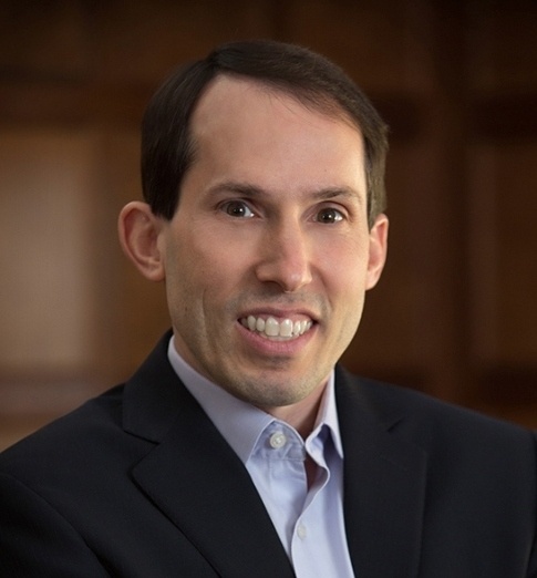
Profile:
Daniel K Sodickson MD PhD is vice chair for research in the Department of Radiology, director of the Bernard and Irene Schwartz Center for Biomedical Imaging, and Professor of Radiology, Physiology and Neuroscience at the NYU School of Medicine in New York City, USA. He also chairs the National Institutes of Health (NIH) study section on biomedical imaging technology. He is credited with founding the field of parallel imaging, which allows distributed detector arrays to gather MR images at previously inaccessible speeds. As a result of his discovery, most MR scanners have parallel imaging hardware and software, and parallel imaging acceleration is used routinely in clinical MRI examinations and research imaging studies worldwide.
27.02.2018