Gender differences in cerebral activation when viewing erotic stimuli in fMRI
By Dr Elke Gizewski, fMRI specialist, Dept. of Diagnostic and Interventional Radiology and Neuroradiology, University Clinic Essen
Functional MRI (fMRI) is a widely used method capable of mapping functional regions of the human cortex in near real time during specific tasks.
Of particular interest is the opportunity to observe secondary cortical responses, activation due to imagined tasks, memory function, time-resolved pathways through cortical regions, and activation in sub-cortical structures. fMRI uses blood oxygenation changes, which can be imaged continuously while functional centres are being stimulated. Image intensity becomes brighter in signal if more oxygenated blood enters the particular brain area. This concept allows the use of blood oxygenation mechanism to image neuronal activation. Visualisation of this effect is accomplished by simple image subtraction, or by comparison of intensity changes as a function of the paradigm application frequency. Using this method, and statistical analysis of every voxel, leads to a functional map.
There is evidence in common sense and psychological studies that men experience more sexual arousal than women, but also that women in mid-luteal phase experience more sexual arousal than women outside this phase. Recently, a few functional brain imaging studies have tackled the issue of gender differences as pertaining to reactions to erotica. The question of whether or not gender differences in reactions to erotica are maintained with women in different cycle phase has not yet been answered from a functional brain imaging perspective.
To examine this issue, fMRI was performed in 22 male and 22 female volunteers. Subjects viewed erotic film excerpts alternating with emotionally neutral excerpts in a standard block-design paradigm. Arousal to erotic stimuli was evaluated using standard rating scales after scanning.
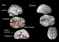
A superior activation for men compared with women in mid-luteal phase was found in the left thalamus, bilateral amygdala, anterior cingulate, bilateral orbitofrontal, bilateral parahippocampal and insular regions (Fig. 1). When comparing men with women scanned during their menses, similar superior activation was found in men. There were no areas of significant superior activation for women compared with men. Comparing women in mid-luteal phase and during their menses, superior activation was revealed for women in mid-luteal phase in the anterior cingulate, left insula, and orbitofrontal cortex (Fig. 2). Sexual arousal was assessed using standard rating scales and did not differ significantly for the men and women during mid-luteal phase, but women in menstrual phase rated significantly lower than the two groups.
Differences in cerebral activity during processing of erotic stimuli can be studied by functional brain imaging. Although hormonal phases of the menstrual cycle influence the specific activation patterns associated with viewing erotic stimuli, gender has the greater influence on differences of cerebral activation patterns. However, there are differences in cerebral activation for women in different cycle times with superior activation in emotion related areas during mid-luteal phase.
These findings may represent a biological representation of differing processing of erotic stimuli, but we cannot answer the question of the underlying mechanisms (genetic predisposition, learning, social factors etc.).
01.03.2006