Article • Gene editing
CRISPR system embeds images in DNA
Report: Mark Nicholls
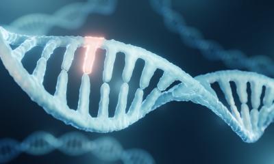
A research team in the United States has developed a revolutionary technique that has encoded an image and short film in living cells. Scientists at the Wyss Institute for Biologically Inspired Engineering and Harvard Medical School (HMS) used CRISPR gene editing to encode the image and film in DNA, using this as a medium to store information and produce a code that relates to the individual pixels of each image. The team hopes to use the technique to create ‘molecular recorders’, an approach ultimately to lead to better methods to generate cells for regenerative therapy, disease modelling and drug testing.
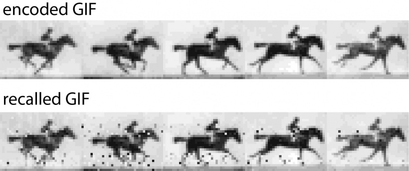
For the research, the HMS group inserted a gif – five frames of a horse galloping - into the DNA of bacteria and then sequenced the bacterial DNA to retrieve the gif and the image, verifying that the microbes had incorporated the data as intended. The images chosen were of a human hand (because it has the type of intricate data the researchers hope to use in future experiments) and a galloping horse by 19th century British photography pioneer Eadweard Muybridge, because it has a timing component that could help to understand better how a cell and its environment may change over time. The team used still and moving images because they represent constrained and clearly defined data sets. The film also gave the bacteria a chance to acquire information frame by frame.
The breakthrough follows work in 2016 when the HMS team built the first molecular recorder based on the CRISPR system. The recorder allows cells to acquire elements of chronologically provided, DNA-encoded information that generate a memory in a bacterium’s genome. The information is stored as an array of sequences in the CRISPR locus and can be recalled and used to reconstruct a timeline of events.
The latest breakthrough confirmed the scientists’ ability to engineer CRISPR system-based technology that enables the chronological recording of digital information in living bacteria. The CRISPR system helps bacteria develop immunity against viruses in their environments.
Capturing viral DNA molecules
Dr Seth Shipman, a postdoctoral fellow in Genetics at HMS, explained that, as a memory of survived infections, it captures viral DNA molecules and generates short ‘spacer’ sequences from them, which it then adds as new elements upstream of previous elements in a growing array located in the bacterial genomes’ CRISPR locus.
The CRISPR-Cas9 protein – a widely used genome-engineering tool – uses this memory to destroy the same viruses when they return, but other parts of the CRISPR system have not so far been much exploited. In this study, the scientists showed that two proteins of the CRISPR system, Cas1 and Cas2, which they had engineered into a molecular recording tool, together with new understanding of the sequence requirements for optimal spacers, enables a significantly scaled-up potential for acquiring memories and depositing them in the genome as information.
‘We designed strategies that essentially translate the digital information contained in each pixel of an image or frame into a DNA code that, with additional sequences, is incorporated into spacers,’ Shipman explained. ‘Each frame thus becomes a collection of spacers. We then provided spacer collections for consecutive frames chronologically to a population of bacteria, which, using Cas1/Cas2 activity, added them to the CRISPR arrays in their genomes. After retrieving all arrays again from the bacterial population by DNA sequencing, we finally could reconstruct all frames of the galloping horse movie and the order in which they appeared.’
Going forward, we’d like to see this work used as the basis for building living biological recording devices that might function as a research or medical diagnostic tool.
Seth Shipman
Effectively, to read the information back, the researchers sequenced the bacterial DNA and used a computer code to unscramble the genetic information. ‘We took on this research because we see the potential for cells to gather information about their own biology and their environment,’ Shipman said. ‘For that to happen, we need a way to capture and store information within a cell while it’s still alive – that’s what we are testing.’
It took researchers three to four years to go from the idea of cells encoding information using the CRISPR-Cas adaptation system to this latest work and it took several days to do the recordings. ‘Going forward,’ Shipman continued, ‘we’d like to see this work used as the basis for building living biological recording devices that might function as a research or medical diagnostic tool.’
Profile:
Seth Shipman PhD is a neuroscientist and postdoctoral fellow in Genetics at Harvard Medical School and a member of a team at the Wyss Institute for Biologically Inspired Engineering. He gained his doctorate, focused on Neuroscience, at the University of California, San Francisco. His key interests lie in genetics and the advancing understanding of brain function.
13.11.2017


