News • Research project
Can new molecular imaging technology guide prostate cancer surgery?

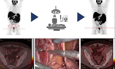
The Netherlands Cancer Institute (NKI) announced that it has received funding from the Dutch Cancer Society (KWF Kankerbestrijding) to test whether a novel molecular imaging technology can guide prostate cancer surgery. The project will evaluate the imaging technology’s ability to detect prostate cancer during surgery, with the aim of performing more accurate removal of cancerous tissue.
Prostate cancer is the most common cancer among men in Europe with over 350,000 diagnosed in Europe every year. Complete removal of malignant tissue during prostate cancer surgery can be challenging, especially in later-stage disease, as it can be difficult to distinguish between cancerous and non-cancerous tissue. Accordingly, there is a crucial need for new technologies to help surgeons visualize cancer in real time during an operation to ensure that the cancer has been completely removed.
If this technique is successful, we will be able to guide the urologist in the most optimal way by combining pre-operative staging with per-operative imaging using the same tracer.
Dr Marcel Stokkel
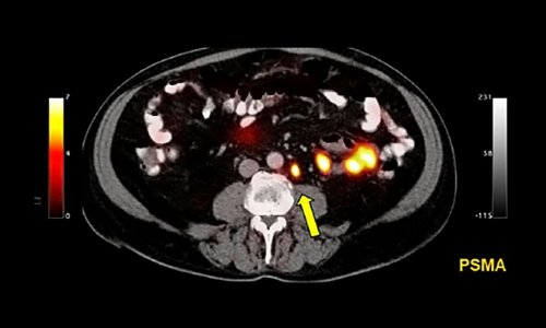
In the joint research project, the Netherlands Cancer Institute, University of Twente and industry partners, Lightpoint Medical and Philips will evaluate a novel molecular imaging technology to image the prostate cancer biomarker Prostate Specific Membrane Antigen (PSMA) during surgery. If successful, the technology will be tested in larger clinical trials with the aim of sparing healthy tissue and reducing recurrence rates in prostate cancer surgery.
Dr Marcel Stokkel, Nuclear Medicine Physician at NKI, commented, “If this technique is successful, we will be able to guide the urologist in the most optimal way by combining pre-operative staging with per-operative imaging using the same tracer. If the resection margins can be assessed with highest accuracy during surgery, we might become able to improve recurrence and survival rates. The collaboration with Lightpoint Medical, Philips and the University of Twente is the most optimal setting to reach this goal and to complete this study.”
“We are delighted to embark on this collaboration with the NKI and Philips. Intraoperative molecular imaging holds enormous potential in prostate cancer surgery. This project will bring the technology a step closer to the patients in need of better treatment options” commented Dr David Tuch, CEO of Lightpoint Medical.
Source: Netherlands Cancer Institute
17.10.2017