Breakthrough
How Dual Source technology is revolutionizing CT
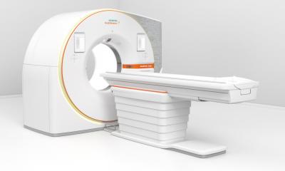
Since launching Somatom Definition in 2005, Siemens has continued to develop Dual Source technology in order to overcome the remaining challenges in computed tomography. This significant development has made it possible to produce diagnostic images of a patient’s beating heart and coronary vessels without having to artificially lower their heart rate, for example. Scanning speeds that were previously unimaginable are now achievable thanks to the temporal resolution of Somatom Definition Flash and Somatom Force Dual Source CT scanners. Increasingly, CT imaging is becoming standard in clinical routine for cardiology. A beating heart can now be scanned in fractions of a second with a radiation dose comparable to conventional X-ray imaging.
From emergency medicine to pediatrics, Dual Source computed tomography (DSCT) has sparked significant progress in numerous other fields of medical imaging – for all patients, regardless of their weight, age, and general state of health.
Technological breakthrough
Siemens built two measuring systems into the CT gantry positioned at 90 degrees from one another in order to achieve higher temporal resolutions and spectral image information. With two X-ray tubes and two detectors in a single system, the foundations for DSCT were laid. The two X-ray tubes and detectors rotate around the patients, acquiring the imaging information twice as fast as single source scanners.
When two X-ray tubes generate radiation at different energy levels – with the electrical voltage of one tube set to 80 kilovolts (kV), and the other to 140 kV, for example – the procedure is called “spectral dual energy imaging”. This technique allows physicians to differentiate between various materials in the body – tissue, bone, implants – with greater precision. It also allows functional parameters, such as the concentration of contrast medium in lungs, heart muscle, or tumors, to be displayed alongside morphological information.
Nowadays, computed tomography has DSCT to thank not only for its significantly higher speed, vastly improved image quality across the entire field of measurement, and greatly increased sensitivity and specificity. DSCT has also eliminated the need for numerous preparation and follow-up care procedures – including the administration of beta-blockers in cardiac CT or the sedation of babies – as well as for breath-holding for thorax imaging. It has enabled perfusion imaging to be successfully integrated into clinical routine, and radiation doses to be drastically reduced.
Fast diagnostics in emergency medicine
When a patient with acute chest pain is brought to the emergency room, time is of the essence. Quick and reliable imaging is key to making a fast and conclusive diagnosis. In order to improve the outcomes of patient treatment and to use the hospital’s resources most efficiently, physicians must perform a triple rule-out and eliminate the three most common causes of chest pain: myocardial infarction, pulmonary embolism, and aortic dissection. A one-stop diagnostic strategy has significant advantages over multiple individual tests and longer monitoring intervals.
For these kinds of trauma cases, where a fast and reliable triple rule-out procedure could prove life-saving, the strengths of DSCT in cardiac and thoracic imaging make a tangible difference.
Siemens DSCT scanners allow physicians to perform diagnostic imaging of the thorax, the coronary vessels, and the entire aorta with one scan and a single administration of contrast medium. Flash mode and Turbo Flash mode enable exceptional imaging quality at lower radiation doses than are required using conventional CT scanners. In the case of pulmonary embolisms in particular, DSCT results in a faster diagnosis and treatment start, since it displays not only the cause – the embolus or several smaller emboli – but also their effect on perfusion in the lungs. In pediatric cases, DSCT has improved the diagnosis of small and distal pulmonary embolisms thanks to its increased specificity and sensitivity. [*]
Dynamic perfusion at dose values of conventional scans
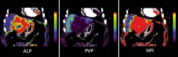
In oncology, therapies can be individually tailored to the particular patient. When it comes to the diagnosis, treatment, and monitoring of tumors – such as in the liver and gastrointestinal tract – individualized therapies demand the most detailed information available on parameters such as blood flow, blood volume, flow time, and permeability.
With Somatom Force, perfusion scans are possible with doses that are no higher than those used for conventional multiphase examinations of the abdomen. The Stellar Infinity detector and the new “Adaptive Dose Shield” dose protection enable up to a 50 percent reduction in the radiation dose for 4D imaging in comparison with other modern CT models – from 30 – 40 down to 12 – 15 millisievert. Somatom Force achieves scan coverage of up to 22 centimeters, enabling the imaging of entire organs. [*]
Functional information on the efficiency of the heart muscle
Coronary CT angiography (CCTA) is a key non-invasive method for detecting coronary artery diseases. If a patient has moderate lesions, however, information about the hemodynamic significance of coronary stenoses is important in deciding whether they would benefit from myocardial revascularization. By performing a CT perfusion examination of the myocardium alongside CCTA, a cardiologist can gain information on blood flow and volume in the heart muscle, and can reliably distinguish between healthy and damaged heart muscle tissue. Following the administration of contrast medium, dynamic CT perfusion imaging acquires several datasets over a period of time in order to precisely determine myocardial perfusion; additional scans or hybrid imaging are then often unnecessary.
Thanks to its high spatial and temporal resolution and large volume coverage, DSCT scanners from Siemens Healthcare is bringing dynamic CT perfusion to clinical routine – leading to improved diagnostic procedures and treatment of coronary lesions.
For a complete list of references, please visit here!
04.05.2016