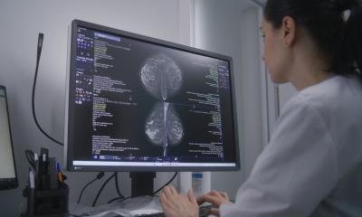
Why low dose really matters in mammography screening
By Dr Jean-Charles Piguet, of ImageRive, Geneva, Switzerland
Although breast cancer, the most common disease affecting women, accounts for over 30% of all the cancers they suffer (far ahead of colon, ovary or lung cancer), breast cancer is responsible for only 1% of cancer related deaths.

In countries with effective screening programmes, over 90% of those diagnosed with breast cancer survive. A decade ago, breast cancer mortality was as high as 50% (50 in every 100 affected women died directly from the disease), which shows the huge improvement in how we treat this disease. However, although therapy has improved, the main reason for the mortality decrease is early diagnosis. This not only changes mortality rates, but also their quality of life: lumpectomy and sentinel node biopsy have a very different impact on a woman compared with a radical mastectomy and subsequent lymphoedema resulting from axillary dissection.
Early detection
As a healthcare provider, for me it is important to acknowledge two very different situations: one with a woman in apparent good health, the other with a symptomatic woman who has felt a lump in her breast. For a symptomatic woman, the clinical situation is straightforward, at least in theory: A mammogram will exclude microcalcifications, show any changes compared with a prior examination, and possibly show a mass or architectural distortion that can be identified as the cause of the symptoms.
For an experienced physician, an ultrasound will clearly distinguish between a typical benign lesion (cyst) and a solid mass. In the latter, the next step is to define the type of mass. Either it is stable and benign, or an ultrasound-guided biopsy under anaesthesia – a painless, inexpensive ten-minute procedure - will provide the histological truth.
In summary, we know how to act with symptomatic women. But we want to find cancers as early as possible, when they are still small, i.e. in asymptomatic women. This means examining a population that is still deemed healthy.
Which technologies are
here to stay?
Screening is now promoted by health authorities in most European and North American countries. The type of modality used, age group invited and frequency of examinations vary from one region to another, but the medical reality is the same.
The vast majority of all women undergoing mammography will have no suspicious lesions: in 1,000 women there are 30 false positives and six to eight cancers are detected, while more than 990 women have undergone a sometimes unpleasant procedure ‘for nothing’. Mammography is costly but, given the benefits, it is very easy to motivate the cost. A more difficult discussion concerns dose. Today, we are exposing a healthy population of women to a lot of unnecessary X-rays to detect a cancer that they, in 99 % of cases, do not have. Again, the benefits clearly outweigh the potential radiation risk, but it is our responsibility to keep the dose as low as possible, which can be achieved in a few ways:
• Reduce the number of exposures
• Increase the interval between examinations
• Reduce the exposure per examination.
The first two methods involve a risk of decreasing cancer detection. The third is the one way to work, provided that exposure reduction does not reduce image quality. Ever since its inception, mammography has been subject to strict government control. Technical limitations of screen film (S/F) mammography produced one set of standards. However, with digital mammography things are no longer that simple. CR tends to further increase dose without improving diagnostic quality, whereas DR significantly reduces the dose. At my clinic, having used all types of technologies and detectors in mammography, the situation for me seems clear: DR will completely replace S/F mammography. CR will also disappear: there is no additional diagnostic benefit compared to S/F and the high dose does not encourage the use of this technology. Also, the poor performance of CR, shown in studies such as DMIST (the Digital Mammographic Imaging Screening Trial), could be widely responsible for the difficulties to demonstrate the obvious superiority of DR. However DR, will be the future.
Three different technologies actually prevail:
1. Indirect conversion with a cesium-silicon sensor and a spatial resolution of 100 microns.
2. Direct conversion with a selenium sensor. There are two suppliers of these sensors, one using 70 microns and the other 80 microns.
3. Photon counting, with a scanning multi-slit detector geometry and pixel size of 50 microns.
Choosing the right system for screening
All vendors obviously argue why their product is superior: from pixel size, DQE, contrast, reliability, workflow, etc. Most radiologists can easily exclude some systems based on image quality. The latter will depend on many factors, including resolution, noise and the algorithms for image processing.
Additional purchasing criteria include ergonomy, workstation features and connectivity/compatibility with different PACS and RIS systems. Additionally, of course, cost is a factor. Although cost is often important, the truth is that, in screening, it should have little bearing on the final decision.
So what about dose?
Too few radiologists think along these lines: if, during the lifetime of my system, I will produce close to half a million exposures of women who are 99% healthy, then is it not my responsibility to truly follow the ALARA* principle? Can I, without reflecting, produce one million mGy (=1’000Gy = 17 full Radio-therapies) when I could, with the same diagnostic quality, reduce this number by 80%? The reason why dose is needed is obvious: a sufficient number of photons must pass through the breast to produce an interpretable image quality.
Additionally, there are at least three factors that allow for a good signal, apart from the dose:
1. DQE: The higher the efficiency of the detector, the lower the dose needed for a good image.
2. Image quality also depends on the scattered radiation. There are two different systems in use to reduce scatter: Anti-scatter bucky grids, and multi-slit collimation of the X-rays before and after the breast, in a scanning system. In terms of scatter rejection, collimation is ten times more efficient, letting through only 3% of scatter compared with 30% by a grid. This obviously translates to a lower dose needed for the same image
quality.
3. Also, the higher the S/N (signal-to-noise ratio), the lower the dose needed to obtain the necessary image quality. Thanks to its photon-counting and detector geometry, the Sectra system that we currently use operates at 50% of the dose or less than all other DR systems, and at about 20% of the dose of CR systems – which are still, against all logic, being sold! Hence the question follows: If the image quality is equivalent, can it be justified to double the necessary dose to women who are 99% healthy? To me, at least, the answer is very simple.
To conclude: In screening mammography over 99% of all mammograms will produce no significant findings. That is the price to be paid for an efficient detection in ‘healthy’ women. The aim is to minimise false negatives and false positives, in other words to have the most sensitive and specific tool possible. DR systems offer good images, in accordance with government regulations, and are obviously better than conventional film mammography. But when one of these DR systems fulfils all the technical and diagnostic requirements, the fact that it uses half the dose compared with any other system should drive towards an unavoidable choice in a screening environment.
* As Low As Reasonably Achievable –
a radiation safety principle and regulatory requirement for all radiation safety programmes, for minimising radiation doses
01.05.2009


