News • Mitosis' structure
Understanding the inside of cancer cells
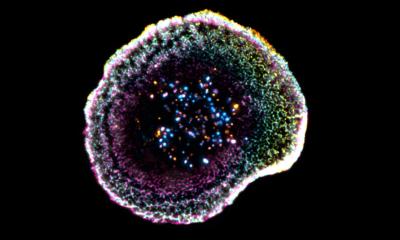
Cell division is an intricately choreographed ballet of proteins and molecules that divide the cell. During mitosis, microtubule-organizing centers (MTOCs) assemble the spindle fibers that separate the copying chromosomes of DNA.
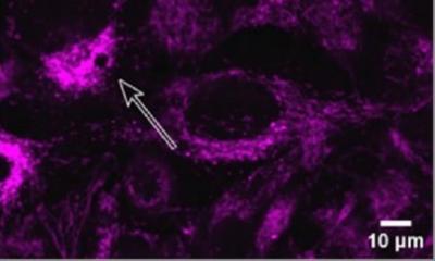
While scientists are familiar with MTOCs’ existence and the role they play in cell division, their actual physical structure remains poorly understood. Shruthi Viswanath, a postdoctoral scholar at the University of California, San Francisco, with a team of researchers, is trying to decipher the molecular architecture of the MTOC. They will present their work during the 62nd Biophysical Society Annual Meeting, held Feb. 17-21, in San Francisco, California. MTOCs are specially designed structures within the cell that create, anchor and stabilize the network of microtubules that act like scaffolding within the cell. More than 1,000 proteins are associated with the MTOC in animal cells, but few of these proteins have been assigned a particular function.
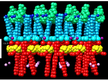
Rather than untangle the complexity of the MTOC, Viswanath focused her research on yeast cells. Within these simpler organisms, the spindle pole body (SPB) functions like the MTOC. Unlike in animal cells, the SPB of yeast contains only 18 proteins and Viswanath has initially focused on five core proteins: Spc110, Spc42, Cnm67, Spc29 and Cmd1.
Using multiple techniques such as structural modeling, X-ray scattering, X-ray crystallography and electron microscopy, Viswanath and her team found that the Spc110 protein provides a greater function in the SPB than originally believed. At first, scientists thought these proteins acted as mere spacers holding pieces of the SPB architecture in place, but now it is believed these proteins may provide a binding surface for this architecture. This information can help understand the function of the human cell equivalent of SPBs called centrosomes. Cancer cells in most forms of cancer reveal abnormalities in the size or structure of centrosomes. Future experiments are necessary to identify the position of other key proteins, like Spc29, a critical protein in the SPB core and to eventually identify its specific function.
Source: Biophysical Society
19.02.2018