Image source: adapted from: Yang L, Wang K, Li W, Scientific Reports 2024 (CC BY 4.0)
Article • Utilizing new strengths, fixing old weaknesses
Ultrasound update for organ imaging
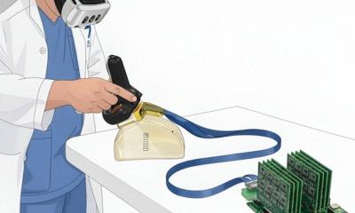
Has organ imaging using ultrasound arrived at the same level as cross-sectional imaging? At the annual conference of the German Society for Internal Medicine (DGIM), PD Dr Corinna Trenker presented new technological developments and their diagnostic significance. Despite numerous innovations such as multiparametric protocols and AI support, she made it clear that the human factor remains one of the greatest strengths of sonography over other modalities.
Article: Wolfgang Behrends
The examination of neck and soft tissue as well as organs in the near-field – testicles, thyroid or breast – has traditionally been one of the strong suits of ultrasound, reported the expert from the Clinic for Haematology, Oncology and Immunology at Marburg University Hospital: ‘With a lateral image resolution of up to 0.1 to 0.2 millimetres, sonography simply exceeds the resolution of CT and MRI.’ This is also reflected in the S3 guidelines – ultrasound is the method of choice for both testicular cancer and the detection of thyroid masses.
[When assessing thoracic wall infiltration in bronchial carcinomas], the decisive factor is the respiratory-dependent fixation of the tumour, and here the sensitivity and specificity of ultrasound is clearly superior to CT imaging
Corinna Trenker
Even though the results of lymph node detection are consistently convincing, the technique has not yet been included in the guidelines, Dr Trenker summarised. Nevertheless, she said that multiparametric imaging using elastography and contrast-enhanced ultrasound (CEUS) is an exciting new tool for assessing lymph node hardening and vascularisation1 – ‘however, this is not relevant for the primary assessment of malignancy,’ the expert added.
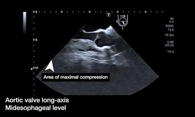
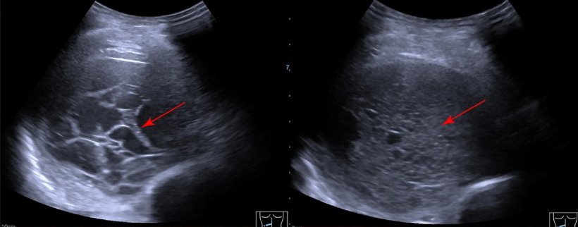
In thoracic examinations, another advantage of ultrasound imaging comes into play: ‘Sonography clearly has the edge because examinations can be performed in real-time,’ explained Dr Trenker. Particularly when assessing thoracic wall infiltration in bronchial carcinomas, ‘the decisive factor is the respiratory-dependent fixation of the tumour, and here the sensitivity and specificity of ultrasound is clearly superior to CT imaging.’2 Ultrasound also remains the method of choice for the detection and differentiation of pleural effusions in 2025. ‘Not only can pleural effusions of just a few millilitres be detected, but they can also be differentiated more accurately,’ said the expert.3
AI opens new possibilities
When it comes to deeper anatomical structures, ultrasound quickly reaches its limits, and the physical limitations of air-filled structures remain unchanged, Dr Trenker pointed out. The high level of examiner dependency also continues to be an issue – but this could change in the future: New publications suggest, for example, that artificial intelligence (AI) could support the traditionally demanding screening for hepatocellular carcinoma (HCC).4 Considering the growing issue of staff shortages, which also affects experienced sonographers, this is a promising development, the expert pointed out – even if there are still a lot of unsolved questions regarding data protection.
Dr Trenker illustrated the amazing diagnostic potential of AI in ultrasound by looking at a recent meta-analysis from China: here, researchers used AI to analyse the sonograms of more than 11,000 breast cancer patients – and, based on this alone, predicted their HER2 receptor status with a high degree of accuracy.5 ‘This is a little hard to grasp, but, at least in initial studies, it is possible. I believe that many more applications in this field will emerge in the future.’
Patient contact: not just ‘nice to have’
Despite the general enthusiasm for technological progress, the expert reminded her audience of one of the key advantages of ultrasound: personal contact with the patient. Studies have shown that this not only creates a more pleasant atmosphere during the examination, but also improves the quality of the diagnosis6, she emphasised. ‘This is where we really have a big advantage over other cross-sectional imaging techniques – and this should not be underestimated,’ she concluded.
References:
- Künzel J, Brandenstein M, Zeman F et al.: Multiparametric Ultrasound of Cervical Lymph Node Metastases in Head and Neck Cancer for Planning Non-Surgical Therapy; Diagnostics (Basel) 2022; https://doi.org/10.3390/diagnostics12081842
- Bandi V, Lunn W, Ernst E et al.: Ultrasound vs. CT in detecting chest wall invasion by tumor: a prospective study; Chest 2008; https://doi.org/10.1378/chest.07-1656
- Yang L, Wang K, Li W, Liu D: Chest ultrasound is better than CT in identifying septated effusion of patients with pleural disease; Scientific Reports 2024; https://doi.org/10.1038/s41598-024-62807-4
- Stefanini B, Giamperoli A, Terzi E, Piscaglia F: Artificial intelligence in Ultrasound: Pearls and pitfalls in 2024; Ultraschall in der Medizin 2024; https://doi.org/10.1055/a-2368-9201
- Fu Y, Zhou J, Li J.: Diagnostic performance of ultrasound-based artificial intelligence for predicting key molecular markers in breast cancer: A systematic review and meta-analysis; PLoS One 2024; https://doi.org/10.1371/journal.pone.0303669
- Gutzeit A, Sartoretti E, Reisinger C et al.: Direct communication between radiologists and patients improves the quality of imaging reports; European Radiology 2021; https://doi.org/10.1007/s00330-021-07933-7
11.11.2025