Toshiba panel of experts
Report: Christian Pruszinsky
Vienna - Toshiba Medical Systems Austria presented a top panel of experts at the ECR to introduce the latest developments in cardiac CT-scanning and dynamic flat-panel technology - the new benchmarks in digital angiography. A further session covered the scientific platform for new uses in ultrasound scanning.
A combination of image amplifier and high resolution CCD camera technology, dynamic flat-panel technology (4.3 lp/mm) is considered top standard for digital and interventional imaging. Special features of the flat panel detector, such as signal interpretation in a single step and minimum pixel size of 150µ x 150µ - suitable for clinical use - deliver more scope for exposure time and they improve image resolution, cut out distortion and reduce the X-ray dose.
P. Peloschek, head of radiology at the Thermenklinikum Baden, expects more detailed imaging and higher resolution from new generation flat panel technology - particularly important, for example, in recognising increasingly smaller details, such as orientation markings on stents. The new technology is expected to offer significant improvements in the diagnosis of abdominal aortic aneurysms, as well as in the examination of peripheral vessel structure in the lower thigh, ankle and neuro-vascular areas.
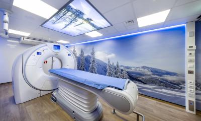
The new Toshiba CT Aquilion 16/cfx, showing 16 slices with 0.5mm per rotation simultaneously, achieves far more detailed coronary images whilst optimising workflow. Combined with specialised, cardiac segmentation software, this achieves a time resolution of up to 50 milliseconds per slice with a rotation time of 0.4 seconds, which means the entire heart can be tomographed with a 16 x 0.5mm selection in 25 seconds. This makes analysis of cardiac anatomy and function possible with only one data set. A special software tool facilitates the automatic reconstruction of axial, sagittal and coronary slices, even with variable parameters, without any extra work. The spiral scan area measures 175cm, so there is no need to move a patient in several examinations.
‘Whether additional features such as panoramic images will become popular depends on the users,’ P. Peloschek pointed out. ‘For progress monitoring of tumour treatment a panoramic view would definitely be a helpful tool.’
He presented a more pragmatic view of contrast medium examinations with ultrasound - currently promoted by ultrasound contrast medium manufacturers to examine inflamed joints, for example. ‘This method is very personnel and cost intensive, like MRI scanning, but documentation is more difficult. The procedure would certainly benefit smaller hospitals, without a MRI scanner, and it may also be of interest for patient examinations in surgeries, but many doctors will probably await further technological developments in this field.’
30.04.2003
- cardiology (790)
- congresses (705)
- CT (617)
- markets (547)
- monitoring (384)
- MRI (848)
- radiation protection (187)
- ultrasound (777)