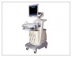
The SonoAce X8

Eliminating Windows-based image protocols, SONOACE in Germany, has produced the SonoAce X8 which, the firm reports, is the first Smart S/W protocol-based ultrasound image processing system.

Photo: The SonoAce X8
‘SonoAce X8 embraces the optimum configuration of hard- and software by using Smart S/W protocol. The equipment not only adapts all the premium functions of existing equipment, e.g. Live 3D, 3DXI, and Full Cardiology Package, but also realizes optimum image performance with Dynamic MR, Full Spectrum Imaging, and SCI (Spatial Compound Imaging).’

The system also can be upgraded in simple steps through a new in-built protocol and  the company adds that the upgraded speed of operation contributes to optimised patient throughput.
Compared with existing ultrasound machines, more accurate and compact data measurements, along with a better reporting function, are promised.

Using a single 17 inch LCD monitor, the system monitors both a patient’s scan and data analysis in real time.

The system generates high picture quality, 1280 x 1024, by using and tracing the best image size for the monitor – the first trial of such a technique in this field, Medison reports.

SonoAce X8 will become commercially available in the first six months of 2007. It is now on show at MEDICA (SONOACE booth: Hall 9, B60) – and later will be at the RSNA in Chicago (later in November).

14.11.2006

More on the subject:

Related articles

Photo

News • Targeted drug activation

Ultrasound-triggered chemotherapy to reduce side-effects

Researchers explore how ultrasound waves can be used to activate chemotherapy drugs only in targeted areas, offering a new path toward safer, more effective cancer treatment.

Photo

News • Multi-lens ultrasound arrays

4D blood flow imaging of whole organs

3D + time: a new 4D ultrasound imaging technique could both improve our understanding of the circulatory system and facilitate the diagnosis of certain blood circulation-related diseases.

Photo

News • Orthopedic ultrasound screening

AI helps spot signs of dysplasia in infant hips

Developmental dysplasia of the hip occurs in 2-4% of infants, but screening via ultrasound is notoriously hard to master. An AI-assisted application could prevent many unnecessary hospital referrals.

Related products

Subscribe to Newsletter