Image source: UT Southwestern
News • Orthopaedic surgery
2D and 3D MRIs add value for ACL surgery
Magnetic resonance imaging (MRI) can reliably establish measurements for anterior cruciate ligament (ACL) “footprints” that are critical to the placement of grafts for reconstruction surgery, researchers from the University of Texas (UT) Southwestern Medical Center report.

Image source: UT Southwestern
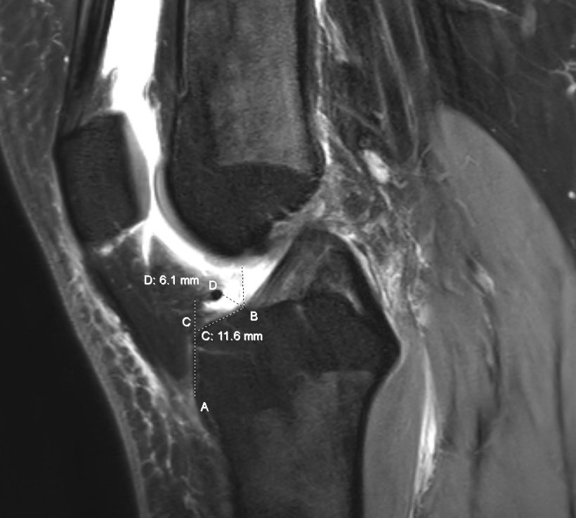
Jay P. Shah, MD, Assistant Professor in the Department of Orthopaedic Surgery at UTSW, and his colleagues used 2- and 3-dimensional magnetic resonance imaging (MRI) to determine these measurements, an approach that had not been explored previously. Their study was published in European Radiology. “This study proves that 2D and 3D MRI is reliable in determining the specifics of the ACL footprint,” said Dr. Shah.
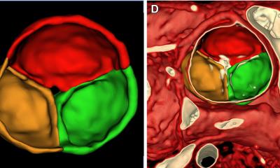
The ACL is a crucial stabilizing ligament of the knee, and an ACL tear is one of the most common ligament injuries with serious consequences in morbidity, mobility, and cost. Among athletes who sustain ACL tears, 37% are not able to return to pre-injury levels. For young patients, ACL tears are often repaired through reconstruction surgery. For successful reconstruction, tunnels must be created in the tibia and femur. The ACL footprint is used to determine where these tunnels should be drilled for graft placement.
We found the mean differences between 2D and 3D measurements of tibia-ACL and intermeniscal ligament-ACL are small, and both 2D and 3D MRI can be reliably used to delineate ACL foot plate anatomy
Jay P. Shah
Anatomical placement of the ACL grafts impacts post-surgery stability and potentially the future risk of arthritis for patients. Incorrect placement of tibial tunnels can lead to reduced rotational stability of the knee. Accurate measurements of ACL footprints will allow surgeons to reliably predict the anatomy and plan for surgery. To determine where to make the tunnels, surgeons typically use arthroscopy, in which a camera connected to a tube is inserted through a small incision to view the joint. However, the locations of the ACL footprints are not always clear arthroscopically.
MRI has been used successfully to observe the structure of the ACL. Dr. Shah and a team of researchers reviewed 2D and 3D knee MRIs from 101 adult patients to measure the tibial-to-ACL and intermeniscal ligament-to-ACL margins. They then compared the measurements to those obtained arthroscopically. “We found the mean differences between 2D and 3D measurements of tibia-ACL and intermeniscal ligament-ACL are small, and both 2D and 3D MRI can be reliably used to delineate ACL foot plate anatomy,” said Dr. Shah.
Establishing ACL footprint measurements using MRI may help reduce surgery times, decrease recovery times, and facilitate better outcomes for patients, added Dr. Shah. Future studies will evaluate the outcomes of ACL reconstruction based on differing footprint measurements.
Source: UT Southwestern
18.11.2022