

Image source: University of Tsukuba
News • Reproducible results
Tau PET brain imaging: standardization to advance disease research
Researchers at University of Tsukuba have developed and released Tomographic Image Preprocessing Using Automated Multi-Reference Estimation for Quantification (TAME-Q), an open-source software package that automatically processes florzolotau (18F) positron emission tomography (PET) images to assess abnormal tau protein accumulation in the brain, a hallmark of several neurodegenerative disorders, including Alzheimer's disease.
The software streamlines PET image analysis and provides researchers with a standardized framework for reproducible data processing and evaluation.
The reseachers published their findings in the journal BMC Medical Imaging.
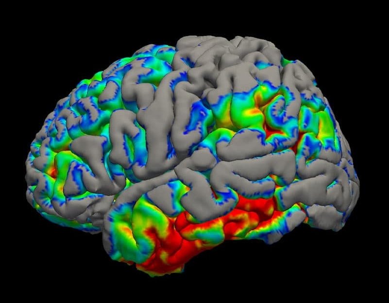
In neurodegenerative disorders, including Alzheimer's disease and progressive supranuclear palsy, abnormal accumulation of tau protein occurs in the brain. Identifying the location and extent of tau accumulation in vivo is crucial for understanding disease mechanisms and progression. PET imaging with florzolotau (18F), a radiotracer that binds to tau aggregates, can visualize tau pathology. The cerebellum is often used as a reference region; however, abnormal tau accumulation also affects the cerebellum in some non-Alzheimer's tauopathies. Therefore, previous studies have proposed estimating reference values from PET signal intensity distribution in gray matter. However, image analysis typically relies on laboratory-specific programs or commercial software, thereby hindering analytical reproducibility and cross-study comparisons.
In this study, the researchers developed and released TAME-Q, software that implements an established semi-quantitative analysis method. It automatically processes PET and structural MRI data, estimates reference values, generates standardized uptake value ratio (SUVR) images, and calculates regional SUVRs as semi-quantitative measures of tracer uptake.
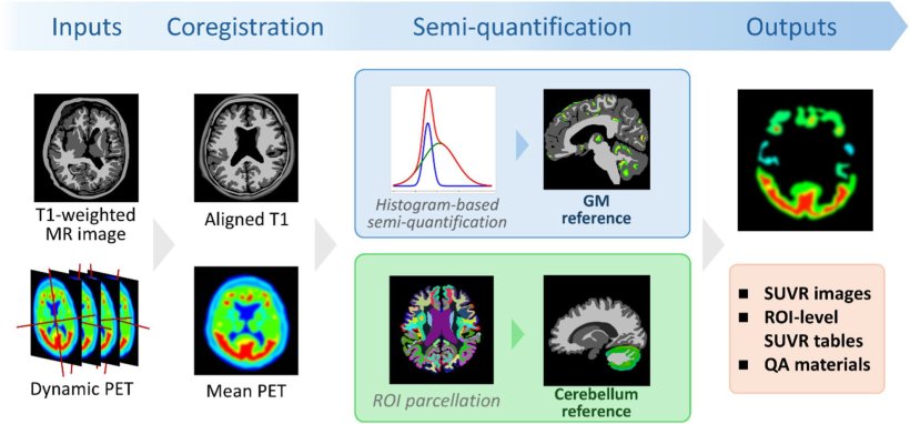
Image source: Nakayama K, Nemoto K, Endo H et al., BMC Medical Imaging 2026 (CC BY 4.0)
The software was evaluated using imaging data from 133 participants: 37 individuals on the Alzheimer's disease continuum, 46 patients with progressive supranuclear palsy—Richardson syndrome, and 50 healthy controls. The automated pipeline successfully processed all datasets, and its measurements were consistent with findings from previous studies of Alzheimer's disease and progressive supranuclear palsy. Additionally, machine learning—derived tau scores calculated from regional SUVRs obtained using TAME-Q correlated with benchmark scores reported in the previous study.
Along with the release of the TAME-Q , the researchers made its execution environment publicly available, allowing analyses to be conducted under the same computational conditions. By reducing the burden of software installation and configuration and providing a transparent, standardized workflow, TAME-Q is expected to support reproducible tau PET research and facilitate future studies of neurodegenerative diseases.
Source: University of Tsukuba
03.11.2026