Synapses recycle proteins for the release of neurotransmitters
Reclaimed proteins enable the fusion of transmitter vesicles with the cell membrane
Neurons communicate via chemical transmitters which they store in the bubble-like synaptic vesicles and release as required. To be able to react reliably to stimulation, neurons must have a certain number of "acutely releasable" vesicles. With the help of a new method, German neuroscientists have now discovered that neurons systematically recycle the protein components necessary for transmitter release and in this way guarantee the reliability of signal transmission in the brain.

If this process is disrupted, the communication between the neurons quickly comes to a standstill and vital processes that rely on the rapid transmission of information, for example seeing or the instant identification of a sound source, become impossible to carry out. (Neuron, November 4, 2010)
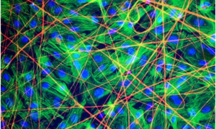
Neurons transmit signals to each other via specialised contacts known as synapses. When a transmitting neuron is excited, it releases chemical transmitters that are discharged by tiny membrane-enclosed vesicles and then reach the recipient cell. The release of the transmitters is carried out through the fusion of the vesicles with the cell membrane - a process that requires the interaction of different protein components in the cell.
Before the transmitter vesicles can fuse with the neuronal membrane they must first be transformed into an active state. The corresponding biochemical process is referred to as priming. During this process, a structure known as a SNARE complex is constructed from protein components that are required for the rapid fusion of the vesicles with the cell membrane.
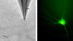
Headed by the Korean neuroscientist JeongSeop Rhee, a group of researchers from the Max Planck Institute of Experimental Medicine in Göttingen have now developed a new method that can be used for the direct measurement of synaptic vesicle priming. The scientists made use of a method that, before now, could only be used for a few special cell types. "Instead of stimulating the neurons electrically, using our measuring system we fill them with a chemically packed signal – containing calcium ions – and then destroy the packaging with a flash of ultraviolet light," explains Rhee. This enabled the scientists to get around many of the complicated processes that normally precede vesicle fusion. They made the process possible by cultivating neurons in Petri dishes on minute islands of just 0.04 square millimetres in size.
With his new method, Rhee and his colleagues Andrea Burgalossi and Sangyong Jung discovered that two protein components, known as SNAPs, play an extremely important role in the recycling of SNARE complexes at synapses. Without SNAPs the recovery of the individual components of SNARE complexes is blocked, and the synapse function also blocked in time.
"We are particularly fascinated by our new method,” says JeongSeop Rhee, "because it provides previously unavailable insights into the mechanisms of transmitter release from synapses." The new information about the priming role of the SNAP proteins is also very important. "A number of pharmaceutical companies are working on processes to influence the priming of synaptic vesicles." If the researchers succeed in regulating this process pharmacologically, it would be possible to develop completely new epilepsy treatments that would avoid many of the side affects associated with the current treatment process.
11.11.2010