Sectra brings PACS power to pathology
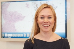
"I can count on my fingers the number of pathologists who work full digitally," said Elin Kindberg who is leading the development of a pathology PACS for Sectra Imaging IT Solutions. Conventional light microscopes continued to be standard tool for examining histo-pathology slides.
Just as the introduction of PACS to radiology departments pushed aside lightboards and led to astounding advances in both reading and reporting, digitization of pathology holds a promise for automating time-consuming tasks, greatly enhancing management of patient files, tracking the chain of production and eliminating the need to go to the glass archive when looking for a previously prepared slide.
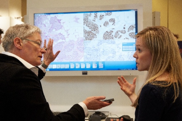
An early realization of this promise was shown when pathologists using digital files to participate in multi-disciplinary tumor boards in Sweden reported meeting times reduced from 90 to 60 minutes."This is not a research project, Sectra PACS for Pathology is a live product that we expect to launch in March, 2015," she said.
Building on the infrasctructure of a radiology PACS is only a starting point, said Kindberg. The front-end interface for pathology analysis and reporting holds very distinct requirements.
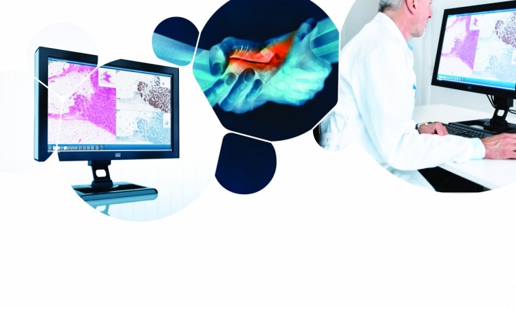
The size of magnified specimen slide images, scanned to convert them from an analog to digital formats, average 500 megabytes per slide. Far too large to be downloaded even at a hospital-based workstation, let alone remote viewing. Streaming to a browser based application is the sensible solution that also builds on the trend toward centralized servers, and especially cloud-based regional archives.
In a demonstration Kindberg called up a patient file using a Google Chrome browser that allows a pathologists to review, quantify, annotate and create a report. The screen can be split into as many as four simultaneous views using a drag-and-drop function. Slides from the same block are automatically registered in all views, tracking with changes to the view in the active split screen.
"Technically it is possible, yes, to stream images to a tablet, but not all the functionality for reporting would be available simultaneously, such as speech recognition and annotation would be frustrating using the tablet interface," she said.
Sectra is strongly focused on the user experience with ongoing studies of pathologists workflow and their ways of working. A 3D control device that operates like a joystick is used to navigate and zoom."Many pathologists complain of having a stiff neck from working on a microscope, they don't want to add carpal tunnel syndrome, or 'mouse arm' to their troubles," said Kindberg.
Unlike radiology where vendors agreed on shared DICOM standrads many years ago, manufacturers of the scanners that digitize blocks of specimen slides have no such agreements and as a result digital pathology data is fragmented among more than 20 proprietary formats.
"We are building a PACS system with vendor neutral standards for viewing and we are open to working with all the vendors," she said.
"Our vision is to take all the good things that come from reviewing and reporting using a microscope, to be sure we have brought them into the digital space, and then accelerate the work with the powerful advantages of PACS," said Kindberg.
09.03.2014