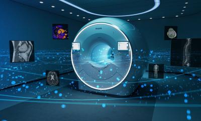
Philips HD15 ultrasound system
Philips has added a new platform to its HD ultrasound products - the HD15 - which aims to provide small hospitals, clinics and private practices with advanced image clarity and broad application support for everyday use.
‘The HD15 brings capabilities that assure user simplicity and productivity to more clinicians in a variety of clinical settings,’ explained Anne LeGrand, senior vice president, Ultrasound, for Philips Healthcare.
‘The system may be used as a primary system for some users, particularly those in emerging markets who require a feature-rich system but may not need all of the features of a high-end ultrasound solution,’ Philips added. Along with workflow improvements, the HD15 capabilities include general imaging, cardiac, vascular and obstetrics/gynaecology applications. Features such as contrast enhanced ultrasound and PureWave transducer technology also allow real-time guidance and evaluation of minimally invasive treatments.
The HD15 includes Philips QLAB quantification software, XRES image processing and PureWave transducer crystal technology. ‘New Microfine EX focusing provides sharper images and improved tissue uniformity throughout the depth of field through application of new dynamic receive lens tuning with five times more focal points than previous generation systems,’ Philips explained. ‘Tissue Specific Imaging presets and iSCAN one button image optimisation can quickly provide clear images with little to no adjustment. A broad suite of configurable patient reports and exam storage options, such as DVD-CD-R/RW, USB drive, and full DICOM capabilities, provide efficient patient data management and colleague or specialist consulting.’
Active native data allows clinicians to manipulate examination parameters and image settings even after a patient has left. ‘Images and Cineloops can receive further investigation by manipulating the original image to see new detail. Live compare allows the clinician to compare a previous exam side-by-side with an active exam in order to see immediately changes in structure or blood flow. This can be particularly helpful in comparing changes in cardiac and vascular anomalies, further documenting changes after interventional procedures, or evaluating foetal development.’
01.09.2008