Image source: SNMMI; from: Schiendler P, Brandt J, Bobe S et al., Journal of Nuclear Medicine 2026
News • FAP-targeted imaging
PET/MRI shows promise in endometriosis detection
A new PET/MRI technique that targets fibroblast activation protein (FAP) overexpressed in endometriosis is more effective at identifying lesions than conventional imaging.
According to new research published in the Journal of Nuclear Medicine, this approach was particularly effective in regions of the pelvis and abdomen that are typically difficult to image. By accurately mapping the extent of endometriosis, FAP-targeted PET/MRI has the potential to enable more personalized management and improve patient outcomes.
Endometriosis is a chronic inflammatory condition characterized by endometrial tissue growing outside of the uterus, affecting 6-10% of women of childbearing age. It is a leading cause of chronic pelvic pain, infertility, and decreased quality of life, posing significant public health and economic challenges. Diagnosis of endometriosis is difficult and is often delayed by four to 12 years.
Recommended article

News • Study shows challenges in primary care
Why diagnosing endometriosis often takes years
Endometriosis affects up to one in ten women and girls of reproductive age, but for many, the path to diagnosis is both long and complex. Now, two new studies explain the diagnostic challenges in primary care.
Until recently, laparoscopy with histologic confirmation was considered the diagnostic gold standard for endometriosis. However, diagnostic laparoscopy is increasingly abandoned because of advancements in diagnostic imaging. Transvaginal ultrasound and MRI are commonly used in diagnosis; however, they may underestimate the extent of disease, particularly in deep infiltrating endometriosis.
“While MRI shows anatomical changes, FAP-targeted PET/MRI highlights tissue remodeling and fibrosis, which are important features of endometriosis,” said Philipp Schindler, MD, radiologist and nuclear medicine diagnostic specialist at the University Hospital Münster in Germany. “In light of the established value of diagnostic MRI, combined FAP-targeted PET/MRI may represent a valuable diagnostic tool for the evaluation of endometriosis by improving diagnostic performance.”
Not only did the combined examination showed substantially more findings suggestive of endometriosis than MRI alone, but these included findings in areas that are often difficult to assess, such as the peritoneum, ovaries, fallopian tubes and locations outside the pelvis
Philipp Schindler
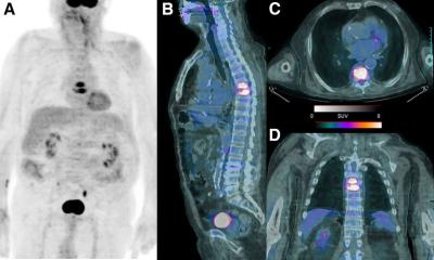
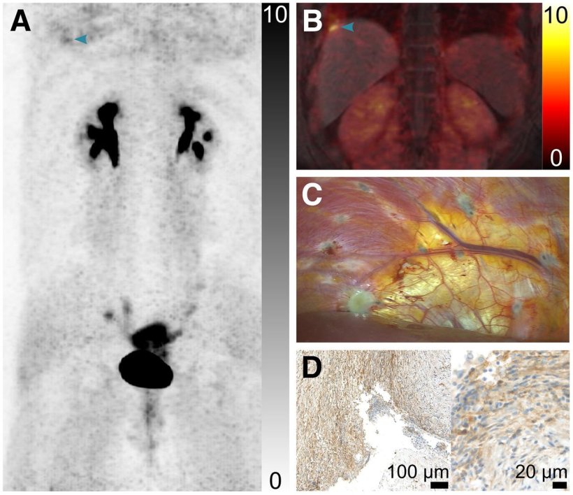
The retrospective study investigated the feasibility of utilizing FAP-targeted PET/MRI to improve endometriosis detection. Participants included 18 premenopausal women: a test cohort of nine patients with suspected or confirmed endometriosis who had undergone FAP-targeted PET/MRI or PET/CT and a reference cohort of nine patients who had undergone FAP-targeted PET/MRI or PET/CT for initial staging of breast cancer. Lesions were assessed using a segment-based analysis and focal radiopharmaceutical uptake was assessed. Clinical assessments and laparoscopic findings, when available, served as reference standards.
In the test cohort with available PET/MRI data, MRI alone identified endometriosis-suspicious findings in 19% of segments. In contrast, combined PET/MRI identified findings in 49% of segments. In the reference group, lesions with focal uptake of the FAP inhibitor radiopharmaceutical outside of the uterus were identified in 14 (13%) of 108 evaluated segments, compared with 44 (41%) of 108 segments in the test group. Where available, PET/MRI findings showed substantial concordance with laparoscopic results.
“Not only did the combined examination showed substantially more findings suggestive of endometriosis than MRI alone, but these included findings in areas that are often difficult to assess, such as the peritoneum, ovaries, fallopian tubes and locations outside the pelvis,” noted Schindler. “If our findings are confirmed, FAP-targeted imaging could establish a new application for molecular imaging in endometriosis, and potentially in other diseases involving tissue remodeling and fibrosis.”
Source: Society of Nuclear Medicine and Molecular Imaging
05.09.2026