© DIgilife – stock.adobe.com
News • AI-assisted platelet imaging
Monitoring blood clotting in real-time
A new imaging technique could personalize heart disease treatment by watching how platelets behave in real time
Researchers from the University of Tokyo have found a way to observe clotting activity in blood as it happens — without needing invasive procedures. Using a new type of microscope and artificial intelligence (AI), their study shows how platelet clumping can be tracked in patients with coronary artery disease (CAD), opening the door to safer, more personalized treatment. Their insights have been published in Nature Communications.
Our technology can help doctors see how each individual’s platelets are behaving in real time. That means treatments could be adjusted to better match each person’s needs
Kazutoshi Hirose
“Platelets play a crucial role in heart disease, especially in CAD, because they are directly involved in forming blood clots,” explained Dr. Kazutoshi Hirose, an assistant professor at the University of Tokyo Hospital and lead author of the study. “To prevent dangerous clots, patients with CAD are often treated with antiplatelet drugs. However, it's still challenging to accurately evaluate how well these drugs are working in each individual, which makes monitoring platelet activity an important goal for both doctors and researchers.”
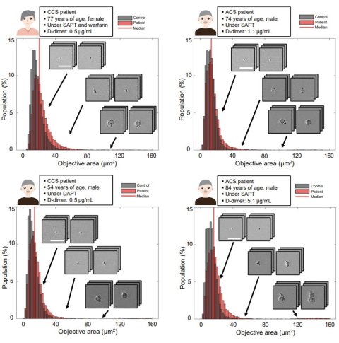
That challenge pushed Hirose and his collaborators to develop a new system for monitoring platelets in motion, using a high-speed optical device and artificial intelligence. “We used an advanced device called a frequency-division multiplexed (FDM) microscope, which works like a super high-speed camera that takes sharp pictures of blood cells in flow,” said co-author Yuqi Zhou, an assistant professor of chemistry at the University of Tokyo. “Just like traffic cameras capture every car on the road, our microscope captures thousands of images of blood cells in motion every second. We then use artificial intelligence to analyze those images. The AI can tell whether it’s looking at a single platelet (like one car), a clump of platelets (like a traffic jam), or even a white blood cell tagging along (like a police car caught in the jam).”

Image source: Hirose K, Kodera S, Nishikawa M et al., Nature Communications 2025 (CC-BY-ND 4.0)
The research team applied this technique to blood samples from over 200 patients. Their images revealed that patients with acute coronary syndrome had more platelet aggregates than those with chronic symptoms — supporting the idea that this technology can track clotting risk in real time. “Part of my scientific curiosity comes from the recent advances in high-speed imaging and artificial intelligence, which have opened up new ways to observe and analyze blood cells in motion,” said Dr. Keisuke Goda, a professor of chemistry at the University of Tokyo who led the research team. “AI can ‘see’ patterns beyond what the human eye can detect.”
One of the most important findings was that a simple blood drawn from the arm — rather than from the heart’s arteries — provided nearly the same information. “Typically, if doctors want to understand what's happening in the arteries, especially the coronary arteries, they need to do invasive procedures, like inserting a catheter through the wrist or groin to collect blood,” said Hirose. “What we found is that just taking a regular blood sample from a vein in the arm can still provide meaningful information about platelet activity in the arteries. That’s exciting because it makes the process much easier, safer and more convenient.”
The long-term hope is that this technology will help doctors better personalize heart disease treatment. “Just like some people need more or less of a painkiller depending on their body, we found that people respond differently to antiplatelet drugs. In fact, some patients are affected by recurrent thrombosis and others are suffering from recurrences of bleeding events even on the same antiplatelet medications,” said Hirose. “Our technology can help doctors see how each individual’s platelets are behaving in real time. That means treatments could be adjusted to better match each person’s needs.”
“Our study shows that even something as small as a blood cell can tell a big story about your health,” Zhou added.
Source: University of Tokyo
15.05.2025


