

Image source: Siemens Healthineers
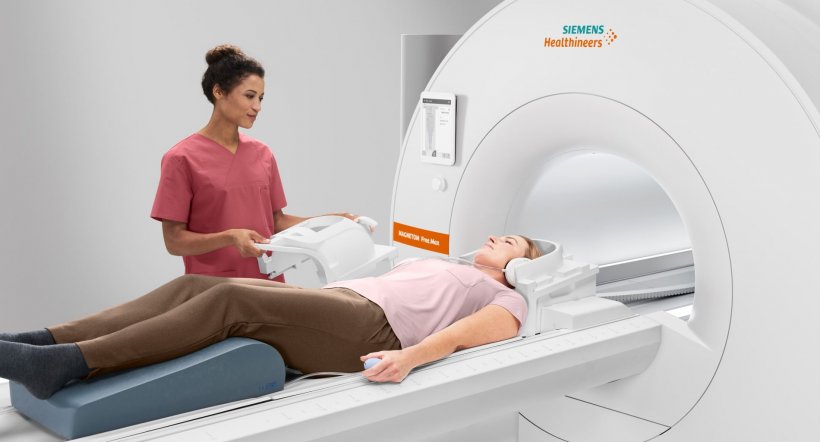
Sponsored • The most compact .55-T full body scanner
Lungs plus A&E MRI scans – no problem
Low, light and with a field strength: 0.55-tesla with added new digital technologies. Indeed, this is a very new class of MRI scanner created by Siemens Healthineers. We interviewed Christiane Bernhardt, Vice President of Magnetic Resonance Marketing & Sales, Business Line MR, at Siemens Healthineers, about the technologies behind this development, plus its advantages and applications.

‘When it came to designing the “High-V MRI” Magnetom Free.Max we specifically opted for 0.55 tesla for a reason. New digital technologies allow us to gain much more information than previously possible with this low field strength,’ explained Christiane Bernhardt, when asked about the new scanner’s field strength. ‘The new field strength offers a range of clinical advantages, including less interference; lower field strength results in fewer implant and susceptibility artefacts. This is an interesting alternative specifically for those patients who need to be scanned at higher field strength due to implants.’
Which other patients does this system suit?
‘The new technology – High-V MRI – allows considerably improved imaging of the lungs compared to today’s 1.5- and 3-tesla scanners. CT may still be the method of choice for visual differentiation between tissue and air in the lungs but, even at low doses, it’s still a system that works with X-rays. Children or patients who need several follow-up examinations at short intervals ideally should not receive this exposure. Our 0.55 tesla scanner is therefore a good alternative for follow-up exams. The first of these scanners has been successfully used for this kind of follow-up in a study on Covid-19 patients at Erlangen University Hospital. The opening of the scanner has a bore of 80cm, which also ensures more patient comfort.’
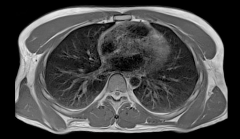
Image courtesy of University Hospital Erlangen
Where would the acquisition of this type of scanner make sense?
‘The Magnetom Free.Max can be considered for the A&E Department, or as a complementary system in the Radiology Department. Out-patient facilities of large hospitals as those often found in the US – so called “urgent care centres” and “hub and spoke centres” – also will certainly benefit from the system. Medical centres and networked surgeries are also potential customers. In addition, we foresee a market for musculoskeletal imaging in orthopaedic practices. And, let us not forget, the new magnet is not only suitable for examinations of the lungs but also for normal, routine examinations of the head and abdomen, for example.’
MRI in an A&E Department – is this rather unusual?
‘Any trauma or acute patient needing ventilation will obviously first be diagnosed via CT. MRI is not the right modality for these cases. But there are some cases in A&E where MRI exams definitely can be justified. Basel University Hospital, where the second Magnetom Free.Max will be installed, plans to use it in the intensive care ward.’
What are the technical innovations?
The Magnetom Free.Max (...) fits through normal width hospital doors and can be transported in a lift, so there is no need to remove walls or for the magnet to be lifted into position by a crane
Christiane Bernhardt
‘The Magnetom Free.Max has new magnet technology and is kept cool with only 0.7l of helium. The cooling system is a completely closed circuit, with no need for a quench pipe. Weighing just over three tons and a transport height of below two metres, this is the lightest and most compact full-body scanner we’ve ever produced. It fits through normal width hospital doors and can be transported in a lift, so there is no need to remove walls or for the magnet to be lifted into position by a crane.’
What about software innovations?
‘One of the new digital technologies is Deep Resolve. This uses a neural network to produce sharper images. The underlying algorithm was trained with pairs of low- and high-resolution images, which allows the reconstruction of higher resolution images by a factor of two. Towards the middle of next year, we will also make Deep Resolve available for 1.5- and 3-tesla scanners with the next version of the software. We now also offer intelligent user control- and guidance for Magnetom Free.Max and MRI examinations, which already proved successful with our CT scanners and X-ray machines.’
What is involved in intelligent control?
The graphically supported operator guidance system called myExam Companion guides the operator through standard exams at the push of a button. If the field of vision for a certain examination is not quite accurate the system will indicate this. It is, of course, also possible to work without this autopilot.
‘As each customer has their preferred protocols, the system – which is also based on artificial intelligence – will initially have to be configured and customised for the specific user. The use of myExam Companion also enables unpractised and inexperienced users to carry out high quality standardised examinations. This means, should it be required (at night, for instance), a radiographer can carry out an MRI scan and send the result to the duty radiologist.
29.11.2020


