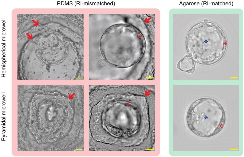
Image source: SPIE; adaped from: Zhao Y, Mc Veigh M, Bellan LM, Bowden AK, Biophotonics Discovery 2026 (CC BY 4.0)
News • Advance in embryo selection
"Invisible" culture dishes improve the odds for IVF
A clearer view for IVF: New "invisible" culture dishes improve embryo selection
Selecting the healthiest embryo is one of the most important steps in in‑vitro fertilization (IVF), yet it remains one of the most uncertain. Roughly 15% of couples worldwide experience infertility, and IVF success rates often remain below 33%. A major challenge is that embryologists must choose a single embryo to implant, relying on what they can see under a microscope. Even small visual details, such as how cells divide or how the embryo’s internal structures form, can signal whether it is likely to lead to a healthy pregnancy. Clear imaging is critical.
With that goal in mind, researchers have explored newer “well‑of‑the‑well” (WOW) dishes, which use small 3D microwells rather than flat dishes. These microwells can support more natural embryo development, but they come with a major drawback: they interfere with optics. Plastics and silicone-based materials commonly used to build the wells bend light differently than the liquid culture medium surrounding the embryo. This mismatch creates blurred regions, warped edges, and visible ridges that obscure fine details. As a result, embryologists must choose between letting embryos grow in a more supportive environment or being able to see them clearly—an impossible compromise in a field where every detail matters.
As reported in Biophotonics Discovery, a team at Vanderbilt University recently developed a promising solution by fabricating WOW dishes from agarose, a hydrogel made mostly of water. Because agarose has nearly the same optical refractive index as the culture medium, light travels through the dish without bending or scattering. In practice, the 3D structure becomes almost optically “invisible,” allowing microscopes to capture sharp, undistorted images.
To test their approach, the researchers compared the new agarose dishes to traditional PDMS versions. They began with optical assessments using tiny microspheres to check resolution and geometric accuracy. In the PDMS dishes, ridges from the manufacturing process visibly warped the image, while in the agarose dishes those ridges nearly disappeared. Details that were previously smudged or interrupted became crisp.
For a more rigorous measurement, the team employed a Shack–Hartmann wavefront sensor, which tracks how light waves change shape as they pass through a material. The sensor revealed that PDMS dishes introduced pronounced and complex distortions, known as high‑order aberrations. In contrast, the agarose dishes produced wavefront patterns almost identical to those seen when imaging through a standard flat petri dish. This confirmed that the hydrogel added almost no optical interference.
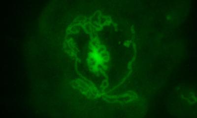
Of course, clear imaging would mean little if embryos could not grow well in the new device. To address this, the team cultured mouse embryos in the agarose dishes and found normal development, matching what is typically seen in established culture systems. Microscopy images showed that structures inside the embryos were sharply resolved, highlighting fine internal features that are important for grading.
With this development, a major barrier to adopting 3D microwell culture has been removed. The agarose-based design allows embryologists to use dishes that promote healthier growth without sacrificing visibility. Combining these strengths could improve the accuracy of embryo selection and, ultimately, contribute to higher pregnancy rates for patients undergoing IVF.
Source: SPIE--International Society for Optics and Photonics
12.02.2026