Image source: University of Eastern Finland
News • Ventricle morphology
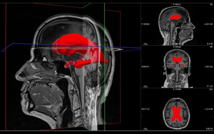
Hydrocephalus: 3D brain marker helps predict shunt surgery success
Researchers at the University of Eastern Finland have identified a new geometric marker of the brain ventricles that could help predict which patients with idiopathic normal pressure hydrocephalus (iNPH) will benefit from shunt surgery.
This is according to a recent study published in Fluids and Barriers of the CNS.
iNPH is a likely underdiagnosed condition in older adults characterised by abnormal gait, urinary urgency or incontinence, and cognitive decline. Unlike most forms of dementia, iNPH stands out because its symptoms can often be improved or even reversed with timely shunt surgery. Neuroimaging is vital for diagnosing iNPH. However, current diagnostic markers are limited and cannot accurately predict which patients will benefit from treatment. As a result, a significant number of patients who undergo surgery may not benefit from it. This highlights the urgent need for more accurate diagnostic tools.
Quantifying the 3D geometry of the brain ventricles can provide important clues about which patients are likely to benefit from surgery
Andrius Penkauskas
In this study, researchers used brain scans from 170 patients with iNPH. Using advanced 3D imaging and machine learning, they quantified and analysed the geometric features of the brain’s lateral ventricles to predict the success of surgical intervention. They found that a geometric marker, asphericity, was strongly associated with better surgical outcomes.
“Our findings suggest that quantifying the 3D geometry of the brain ventricles can provide important clues about which patients are likely to benefit from surgery,” said the study’s lead author, Andrius Penkauskas. “This advancement brings us a step closer to better identifying iNPH patients who will truly benefit from complex brain surgery and sparing many from the suffering of ineffective treatment.”
This research project has received funding from the European Union’s Horizon 2020 research and innovation programme under the Marie Skłodowska-Curie Actions.
Source: University of Eastern Finland
22.04.2026