Image source: Shimadzu
Sponsored • DBS PEth analysis
Fully automated and hematoctrit corrected phosphatidylethanol analysis
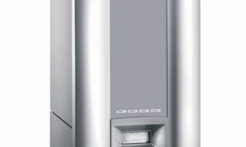
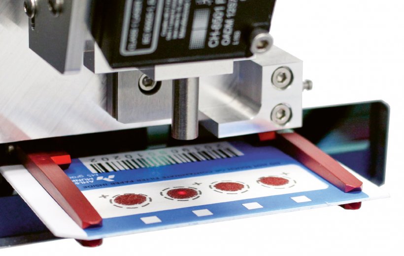
The Swiss-based CAMAG DBS Laboratory in collaboration with the Institute of Forensic Medicine in Bern, Switzerland, has developed a novel approach for the fully automated analysis of the direct alcohol marker phosphatidylethanol (PEth) in dried blood spots (DBS). The use of a DBS autosampler with an embedded hematocrit (HCT) scanner combined with an LC-MS system permits analysis of large sample volumes and correcting for the red blood cell volume percentage, without any manual sample preparation.
With a total analysis duration of only 5 minutes per DBS sample, including photo documentation of the DBS card before and after the extraction, hematocrit measurement, and internal standard spray application, the developers believe that the novel fully automated workflow is quicker, easier and cheaper than current PEth testing routines. Manual steps such as cutting DBS, adding extraction solvent and internal standard, mixing and shaking, centrifugation, evaporation and reconstitution of the sample become obsolete. Furthermore, the use of DBS cards permits sending the specimen in an envelope to a centralized laboratory without cooling.
Dr Marc Luginbühl, principal scientist at the CAMAG DBS laboratory, says: “The novel workflow has been validated. In comparison to liquid blood extraction and manual DBS extraction it shows excellent reproducibility. Additionally, with the latest implementation of a HCT scanner to correct for the red blood cell volume percentage, we have made a crucial step forward. As PEth is mainly present within the red blood cell fraction, the sample’s hematocrit should be taken into account when doing PEth analysis, as it can significantly influence the outcome. Using the instrumental setup presented here, this becomes accessible in a straightforward and fully automated way for the first time.”
What is PEth analysis needed for?

Phosphatidylethanols are formed as abnormal phospholipids within the human body when ethanol is present. PEth itself is not a single molecule, but rather a group of molecules that differ from each other in their fatty acid chain composition. As PEth accumulates in the red blood cells, which prolongs its window of detection, it became popular as a direct alcohol biomarker. Thereby, PEth’s strength lies in recognizing the drinking habits during the past two to four weeks, rather than the detection of a single drinking event with limited quantities of alcohol.
PEth 16:0 / 18:1 and PEth 16:0 / 18:2 are the two most abundant PEth homologs in human blood and are often measured together. The concentration of PEth 16:0 / 18:1 is frequently used to classify alcohol consumption behavior based on two threshold concentrations: a lower threshold is used to distinguish between light or no alcohol consumption and is usually set at 20 ng / mL (~0.03 μmol / L). An upper threshold is used to distinguish between substantial alcohol consumption and heavy alcohol consumption and is set at ~200 ng/mL (~0.3 μmol / L).
The analysis of PEth is applied in the follow-up of alcohol-impaired drivers, diagnosis and treatment of alcohol use disorders, newborn screening for fetal alcohol syndrome, organ transplantation and in the security environment. Compared to other direct or indirect alcohol markers (EtG, CDT, or GGT) PEth was found to have better sensitivity and specificity in various studies.
Fully automated PEth analysis
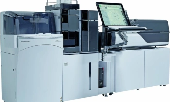
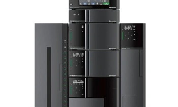
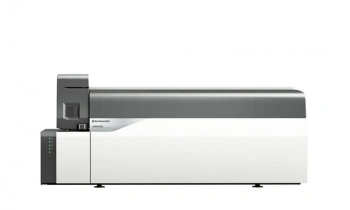
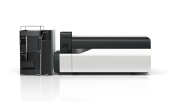
The combination of a CAMAG DBS autosampler with a Shimadzu 8050 LC-MS system permits a hyphenated fully automated DBS-LC-MS / MS workflow. Thereby, the DBS specimen is eluted onto a short trapping column (Polar RP, 20 mm), separated on a longer analytical column (C5, 50 mm), and subsequently detected using electrospray ionization tandem mass spectrometry.
The use of a double-column system has the major advantage that a solid-phase extraction like purification of the target analytes is achieved, which reduces matrix effects, co-elution of disturbances and overall system contamination. In contrast to manual, passive extraction of DBS using a tube and a mixer at ambient pressure, the automated system performs an active extraction by constantly pumping fresh extraction solvent through the DBS.
After the extraction of each DBS sample, the extraction cell of the autosampler is washed with a selection of three different wash solutions. By using an additional six-port valve installed on the Shimadzu LC-MS system, seamless switching between the CAMAG DBS autosampler and the standard vial autosampler is possible without reconnecting any capillaries.
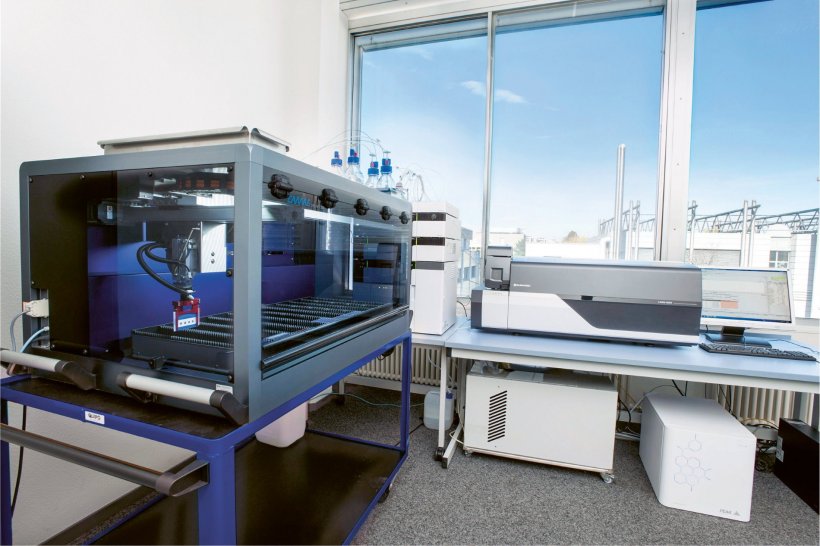
Image source: Shimadzu
Why use dried blood spots?
PEth showed limited stability in liquid blood unless stored at – 80°C. It was observed that the use of dried blood microsamples increases the sample stability: using DBS, the post-sampling formation and degradation of PEth can be prevented and the stability of the samples is improved, as any enzyme activity is stopped during the DBS’s drying process. Overall, the collection of DBS microsamples is easier, less invasive and cheaper than conventional whole blood sampling, as samples can be handled, shipped and stored more cost efficiently over a prolonged time. In addition, specimen collection can be done by untrained persons, including the study subjects themselves and in remote settings.
How does the DBS hematocrit measurement work?
“What we developed and implemented for PEth analysis”, Marc explained, “is a way of obtaining the hematocrit from already dried samples in a fully automated fashion, thus making it possible to measure hundreds of DBS samples in a non-destructive way.”
The automated hematocrit detection is based on a patented reflectance measurement setup and is not influenced by humidity, temperature or the age of the DBS. During the measurement, a probe illuminates the DBS and measures its reflectance at a very specific wavelength. A built-in laser allows controlling and adjusting of the distance between the probe and the DBS.
As the signal measured is proportional to the hematocrit of the sample, the red blood cell volume percentage can be calculated based on a pre-recorded calibration curve. Thereby, the HCT scanner allows normalizing for any known HCT related DBS effect such as the HCT area bias, the HCT recovery bias, or HCT related analyte properties. Furthermore, it is possible to normalize the analytical result to a selected HCT value. The system is ideally suited for the reliable, quantitative analysis of non-volumetric DBS samples.
Profile:
Dr Marc Luginbühl is the principal scientist of the CAMAG DBS Laboratory, which develops automated dried blood spot solutions. His specific areas of expertise lie in the assessment and measurement of biomarkers using microsampling strategies with a passion for drug of abuse screening and alcohol biomarker analysis. With a PhD in Chemistry and Molecular Sciences, he is a member of The Society of Forensic Toxicologists (TIAFT) and the founder of The Society of Phosphatidylethanol Research (PEth-NET).
09.12.2021


