Image source: Materialise
Interview • Personalised implants in CMF surgery
Digital planning in skull reconstruction: from benefit to necessity
Whenever cranioplasty is discussed, the conversation usually centres on the implant material: titanium or PEEK. However, for Prof. Thomas Schouman, a CMF surgeon at the AP-HP Pitié-Salpêtrière Hospital in Paris, and Maarten Zandbergen, Market Manager for Cranio-Maxillofacial Solutions at Materialise, predictability begins much earlier – with digital planning, long before the patient enters the operating theatre.
Interview: Wolfgang Behrends
HiE: How were complex skull defects managed before personalised implants became available, and what has changed most fundamentally as a result of patient-specific implants and instruments?
Prof. Thomas Schouman: ‘Before personalised implants, complex skull defects were mainly reconstructed with bone grafts taken from the patient. This could be a split calvarial graft, a graft from the iliac bone, or sometimes a rib graft.
The first issue is that you need to harvest bone somewhere else, so the patient has a second surgical site. And when we are dealing with large skull defects, we often need quite a large amount of bone – which we then have to shape manually. There is uncertainty about the healing of the graft, and in some cases the graft can be lost.
Another material that has been used a lot, and is still used quite often, is PMMA – polymethyl methacrylate. It is inexpensive and fairly easy to use, but of course, the shape is not always perfect. It can also become infected, especially if it is close to the sinuses.
Personalised implants changed things quite fundamentally because the implant is designed to fit the defect before the surgery. This eliminates the need to harvest bone, and shaping the reconstruction during the operation. The surgery becomes much more straightforward. It is shorter, easier, and more predictable.
In my own practice, since patient-specific implants have been available, I do not recall using autologous bone grafts anymore for significant skull reconstruction. For me, in large skull defects, it was definitely a game changer.’
Maarten Zandbergen: 'What we saw from our side, working with cranial surgeons since the late 1990s, is that they shaped the reconstruction at the table in the OR: autologous bone where the defect allowed it, or titanium mesh and bone cement shaped by hand.
What changed is that the reconstruction is now designed beforehand on the patient’s own anatomy, and the surgeon reviews and approves it before the operation. This has taken away a lot of uncertainty and increased predictability.’
How exactly is digital planning changing the way surgical teams approach complex skull defects?
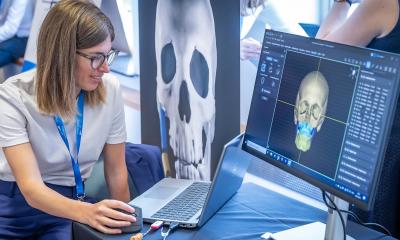
Zandbergen: ‘It changes the order of the work. The CT scan becomes a virtual 3D model that the case is built on, rather than an image to look at. The defect gets accurately measured and the reconstruction is designed against the healthy side, or against a reference anatomy when no healthy side is left.
The 3D model is not only a visualization tool but also a powerful aid in treatment decision-making and a design environment where clinical requirements are translated into a feasible implant.’
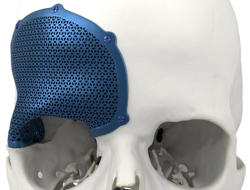
Schouman: ‘The practical effect is that we move part of the complexity before surgery. Usually, the engineers propose a first design, showing the general volume and shape of the implant. We check that it meets the clinical requirements – whether the implant fills the defect properly, whether the position of the fixation screws is appropriate, and whether the implant can be inserted through the surgical approach we plan to use. Then they move to the final design, with the details of the implant, for example, the mesh if it is a titanium implant. Sometimes the implant is very large, and depending on the access, it may be difficult to insert it in one piece. In those cases, we may decide to split the implant into two parts.
A lot of the work is on the engineering side. Planning remains very important because it ensures that the implant aligns with the defect, the patient’s anatomy, and the surgical approach. But with a personalised implant, the main impact is that the reconstruction is already prepared before entering the operating room.’
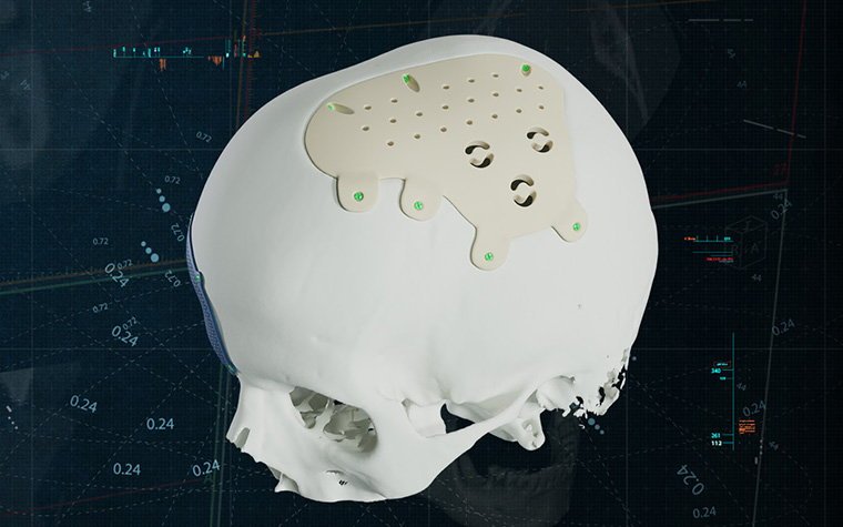
Image source: Materialise
What role does the choice of material (titanium or PEEK) play?
Schouman: ‘The choice of material is very important. It depends on the type of defect, the location, the condition of the patient, and the risks we want to avoid.
Porous titanium implants can integrate very well with the bone. But this can also be a drawback. If you need to remove the implant later, it can be very difficult to take out because of this integration
Thomas Schouman
For example, when the defect is close to the paranasal sinuses, close to the orbit or the nose, so especially in the anterior part of the skull, the risk of infection is higher. In these cases, I would rather use titanium.
Porous titanium implants can integrate very well with the bone. But this can also be a drawback. If you need to remove the implant later, it can be very difficult to take out because of this integration.
There are also imaging issues. Titanium is very visible on CT scans, and it can create artifacts. It does not prevent MRI, but it can disturb the images.
So there is no perfect material. Each one has advantages and disadvantages. The important thing is to choose the material according to the clinical situation.
There is also a practical aspect, at least in France, which is reimbursement. PEEK and ceramic implants are reimbursed, whereas titanium implants are not. That has an impact on the strategy.’
Zandbergen: 'Titanium 3D printed implants gives mechanical strength and design freedom, including porous structures that can support bone ongrowth, and it fixes reliably with self-drilling screws.
PEEK is radiolucent, so follow-up imaging stays clean. It is light, and it can be adjusted intraoperatively for screw fixation. It suits cranial and orbital reconstruction, facial contour restoration and onlay applications, with limited bone ongrowth.
Neither material is the better one. What matters is that the choice can be made on a case-by-case basis, so the surgeon can consider multiple options, taking into account the patient’s needs.
From our perspective, the important point is that material choice is not separate from planning. Titanium and PEEK behave differently, so the design has to account for the material from the start, including information about fixation, thickness, imaging needs, and how the implant will be handled in the OR.’
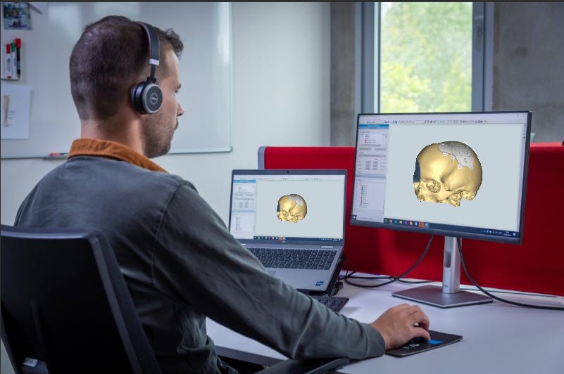
Image source: Materialise
What are the benefits of close collaboration between imaging, planning, implant design, manufacturing, and clinical practice?
Schouman: ‘I would not say it is only a benefit – it is a necessity.
There has to be a close dialogue between the clinicians and the engineers to make sure that the surgical approach and the implant are compatible. There is nothing worse than starting a surgery and realising that you cannot insert the implant through the approach you planned.
An important point is soft tissue closure: After a skull defect, especially if it has been present for some time, the soft tissues can retract. Usually the defect creates a concavity, and the reconstruction changes this into a convex shape again – so we are adding volume under the skin. Whether the wound can be closed after inserting the implant, depends on the volume of the reconstruction, but also on the quality of the soft tissues, and the patient’s history – previous surgeries, scar tissue, radiotherapy. In some cases, we may need a soft tissue flap to have enough coverage for the implant.’
In CMF surgery, every patient is different and every step depends on the one before. Holding those steps together on a single plan puts predictability in the hands of the surgeon and the team around them
Maarten Zandbergen
Zandbergen: ‘Those five steps only work together if something holds them together, and for us that is the 3D plan. It is built from the scan, it is what the surgeon reviews and approves, it defines the implant design, and it drives production. Supported by an online case management platform, it ensures that everyone works and collaborates on the same file.
The benefit is increased predictability. In CMF surgery, every patient is different and every step depends on the one before. Holding those steps together on a single plan puts predictability in the hands of the surgeon and the team around them.’
Recommended article
Article • Technology applications
Medical 3D printing: from niche to mainstream
From personalized medical guides and implants to advanced surgical planning solutions: 3D printing and visualization has seen considerable growth over the past years and is already making a significant impact in healthcare. AI, cloud, and virtual/augmented reality technologies show promise to further advance the number of applications.
Where do you see the greatest advances in complex skull reconstruction in the coming years?
Schouman: ‘For me, the technology for designing personalised implants is already highly developed and very efficient. The main limitations now are more related to the materials: We need materials that have the right balance between strength, integration, infection resistance, imaging compatibility, and removability.
Whether bioresorbable implants will become relevant for large cranial reconstruction. I really don't know.
There is also the question of reimbursement. Broader reimbursement of patient-specific implants, including titanium implants, would help both patients and surgeons.’
Zandbergen: ‘We see it evolving in three directions. And none of them is a new implant.
First is about reach: A 3D plan and personalized implant are becoming standard practice for complex cases, not yet for every case, because planning and design take expert time. Automation can support routine preparation, while expert review, validated workflows and surgeon approval remains central.
The second concerns soft tissue prediction: Better use of data that has already been acquired, and planning that accounts for soft tissue rather than bone alone. Bone is what we can plan with precision today, while the result the patient sees is soft tissue.
Finally, access: Personalized implants and 3D planning are still limited by reimbursement across much of Europe; with better reimbursement systems in place, more patients will be able to benefit from personalization.’
Profiles:
Prof. Thomas Schouman is a Professor of Maxillofacial Surgery at Sorbonne University's Faculty of Medicine and a senior surgeon at AP-HP Pitié-Salpêtrière Hospital in Paris. A pioneer in personalized cranio-maxillofacial surgery, Prof. Schouman has been at the forefront of integrating patient-specific 3D-printed solutions into clinical practice for over a decade.
Maarten Zandbergen is Market Manager for Cranio-Maxillofacial (CMF) solutions at Materialise, where he leads the company’s global CMF portfolio. Since joining Materialise in 2007, he has developed deep expertise in medical image processing, 3D planning, and patient-specific solutions for cranio-maxillofacial surgery. He collaborates with clinical and industry partners worldwide to advance innovative digital and 3D-printed applications in healthcare. Maarten holds a Master’s degree in Mechanical Engineering, with a specialization in Biomedical Engineering.
17.09.2026